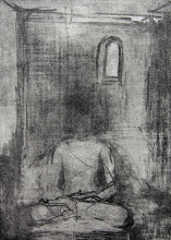
Sunday, December 31, 2006

Painting post - last one for this year - and a happy new year on that note:


This did not come out as well as I wished it would, but this oil titled 'Hazy Shades of Winter' is 48 inches by 36 inches. I finished it in about four days and maybe that explains why parts of it are a bit sloppy (I was not the happiest with this one)... You tell me....

Friday, December 29, 2006

When opposites become apposite...


In my view, science is the ultimate unifier - looking relentlessly for universal truths that explain phenomena with simple clarifying concepts. Art on the other hand is in the other extreme - the epitome of self expression, uniqueness and individuality. It is indeed refreshing when the both come together. OK, what am I talking about now…

These two worlds came together in a refreshing fusion this week at Princeton University.

Art of Science asked the Princeton University community to submit images including videos and audio produced in the course of research or incorporating tools and concepts from science. Here are the results.
Quite amazing, I must say!!

Thursday, December 28, 2006

Sobering column that appears everyday in the New York Times (for the last four years) this was from today's Times:


You know after a while a lot of this becomes just statistic. I usually scan the names, remember the dead people (soldiers and the uncounted civilians) and move on - but this time I was struck by the fact that all people listed dead above were in their 20's.

Tuesday, December 26, 2006

The teen brain - a work in progress and driving implications :


An overwhelming amount of research is slowly bringing to light the fact that the teen brain is very much a work in progress and a lot of the 'neuronal sculpting and pruning' that takes place as the child matures through the teen years is incomplete at least until the age of 21 to 22... I remember as a teen (about 20 years back), I used to sneak into a lower shelf in my family cupboard and 'help myself' to money kept there to to buy cool jeans. For some reason my father did not confront me, but one day the goose that laid the golden eggs stopped replenishing itself and I often wondered if my father 'noticed'. I also remember sneaking out on my father’s motorcycle a couple of times without his knowledge and without any sort of a valid license to drive. I was 15 then. Looking back in retrospect, it seems stupid and unreasonable to assume that my father would not 'notice'. Research can now conclusively tell us that the teen brain is undergoing a huge amount of structural changes. In fact the following have been conclusively proved:

1. The first areas to mature (e.g., extreme front and back of the brain) are those with the most basic functions, such as processing the senses and movement.
2. Areas involved in spatial orientation and language (parietal lobes) follow.
3. Areas with more advanced functions -- integrating information from the senses, reasoning and other "executive" functions (prefrontal cortex) - mature last.

The actions outlined in 3 above takes place post puberty and extends over 8-10 years (until they are 21 or 22)... In fact actions that are directed by the pre-frontal cortex involve critical integrative functions like organize plans, generate ideas, set priorities, form strategies, control impulses, and allocate attention. In retrospect all of this was what I lacked when I helped myself to get the jeans and drove outside on my motorcycle in unbridled frivolity.

In fact a couple of months back, there was the case of a teen who texted her suicide notes as she drove her Mercedes into a oncoming Daewoo. The mother of three in the Daewoo died as a result while the teen survived. Highly irrational behavior like this can be rationalized a bit more when we understand that Louise Egan Brunstad's (later charged with murder as an adult) neuronal connections were not sculpted or pruned enough for her to think or plan or strategize her next course of actions in light of her current conditions. Of course, this opens up a bunch of ethical arguments again - do not blame the child, blame the brain - but nonetheless brings to light the fact that we need to pay closer attention to teen brains in formative modes and maybe legislate that actions that require a surprisingly large amount of planning and thinking - like driving for example should maybe be the purview of 21 year olds instead of 16-18 year old...

Of course, some of this might be a little extreme, but Louise’s case above and a bunch of other similar cases may have a case in point.

Related papers of interest:

The Adolescent Brain: A Work in Progress - A good article on changes going on in the teen brain with a focus on preventing teen pregnancy

What’s Going On In There? How the Brain and Mind Develop in the First Five Years of Life By Lise Eliot, Ph.D.

What Makes Teens Tick? - Article in TIME on the teen brain...

Cruel and Unusual Punishment: The Juvenile Death Penalty Adolescence, Brain Development and Legal Culpability - A good flyer put out by the American Bar Association that talks about law and the teen brain and the ramifications

Neuronal Regulation: A Mechanism For Synaptic Pruning During Brain Maturation by Gal Chechik and Isaac Meilijson
- a little mathematical, but a good read nevertheless

Are Teens Driving Safer? By Pilar S. Marin and Brett V. Brown, Ph.D, Child Trends

The First Three Years of Life and the Early Adolescent: Influences of Biology and Behavior - Implications for Child Rearing by Donald E. Greydanus, MD; Helen D. Pratt, PhD; Dilip R. Patel, MD - A CME review article

Teen driving - should states impose tougher restrictions?

Saturday, December 23, 2006

Brains filling in or blanking out?

What you see is not reality in this really impressive optical illusion...

http://www.patmedia.net/marklevinson/cool/cool_illusion.html

"If your eyes follow the movement of the rotating pink dot, you will only see one color, pink. If you stare at the black + in the center, the moving dot turns to green. Now, concentrate on the black + in the center of the picture. After a short period of time, all the pink dots will slowly disappear, and you will only see a green dot rotating if you're lucky! It's amazing how our brain works. There really is no green dot, and the pink ones really don't disappear. This should be proof enough, we don't always see what we think we see."

The following site gives a lot more optical illusions that fool our brains if you are really into this kind of stuff... http://www.michaelbach.de/ot/index.html

Tuesday, December 19, 2006

An aside on ethical dilemmas in brain research:

As research deepens our understanding of the human brain, it is becoming clearer that the distinctions between abnormal and normal brains and concomitant functions are getting more and more blurred. In many cases we are slowly proving that abnormal behavior manifested is due to either inbuilt genetic makeup or genetic changes forced due to conditioning of the human brain in particular environmental settings.

Distinctions between mental or psychiatric diseases and neurological disorders are lost as science progresses towards a more exact understanding of neurological phenomena and unifying explanations that seem to reduce the stigmas associated with psychiatric diseases. On the one hand it empowers the patient in the sense that patients seem to have a better understating of exactly why they are behaving in a certain fashion (abnormal to many but seemingly normal to the patient and the neurologist) down to the molecular detail. This gives the patient a new sense of confidence and in some cases causes reversal of the stated condition (a case of knowing the ailment modifying the behavior of the individual).

On the flip side, we now run the risk of having criminally violent individuals armed with new knowledge of what goes on inside their heads down to the molecular level portraying themselves as mere puppets of the neural machinery that they are endowed with and shaped by the environment. The extreme case of a newly-wed filing a £3.5 million compensation claim against employers, stating that his marriage was ruined because his sex drive spiraled out of control after he injured his head at work is a case in point.
As pointed out in numerous papers at the end of this post, research is revealing patterns of neural activity and gene lines that may be responsible for criminally violent behavior. How do we deal with individuals like this - blame ostracize the individual who committed the crime or blame the environment that the individual was placed in that modified his neural genetic machinery to make the person behave in a 'abnormal (read criminally violent)' fashion..? What about the future when neurologists can predict the predisposition of certain individuals to 'abnormal' behavior based on certain ‘environmental markers’ that could change their neural machinery in socially inappropriate ways? When would society decide to intervene and make changes in an individual’s life based on 'prescient' information about the nature of ones evolving neural machinery..
This is but of a host of minefields that will undoubtedly be uncovered as humankind goes deeper into unraveling the mysteries of what lies within our heads...


Monday, December 18, 2006

Painting post:

OK, I managed to finish this one over the weekend, took about three weeks to complete, but I think it was well worth the effort. Again, this canvas is one of my standard sizes (48 inches by 36 inches) and the technique consists of juxtaposing solid oil tones against opposing contrasting hues. I am satisfied with what I achieved this time around although I am still refining the technique.
The painting is titled 'Buy Me Bring Me Take Me'.

Thursday, December 14, 2006

Congenital AVMs and the United States Senate:

Majority control of the United States Senate lies a bit in balance with one of the senators of the leading party in hospital after suffering a brain hemorrhage. I thought this might be a good time to focus on the nature of the condition that led to the hemorrhaging in the senator’s brain. The senator suffered from what is called a "congenital arteriovenous malformation". Even if the name sounds a little foreboding, this is relatively simple to understand.

Normally, arteries carry blood containing oxygen from the heart to the brain, and veins carry blood with less oxygen away from the brain and back to the heart. When an arteriovenous malformation (AVM) occurs, a tangle of blood vessels in the brain or on its surface bypasses normal brain tissue and directly diverts blood from the arteries to the veins. It is estimated that about one in 200–500 people may have an AVM and more common in males than women. Brain AVMs are usually congenital but not hereditary (meaning you may be born with one but will not pass it onto your children)...

As a brain AVM contains abnormal (“weakened”) blood vessels, they sometimes dilate over time and may eventually burst from the high pressure of blood flow from the arteries causing bleeding into the brain. The bursting may be precipitated by several factors that include exercise, stress or sometimes something as common as a sudden stop while riding in a car…

Most AVMs are detected on either a computed tomography (CT) brain scan or with a magnetic resonance imaging (MRI) brain scan. These AVMs can be surgically removed if bleeding occurs. The other ways of treating this would be to 'scar' part of the blood vessel supplying the AVM (using a technique called stereotactic radiosurgery) and allowing the AVM to 'clot off'. There are other advanced ways of treating this inclusing the usage of catheters inside the blood vessels and deploying certain specialized materials to block off the blood flowing into the AVM. Not too sure which one of this was performed on the senator..

It is interesting to note that the senator showed signs of speech slurring and had to be taken to the hospital following that. This would indicate that the AVM was somewhere in the left anterior parietotemporal lobe (just my layman theory) ;-)

Wednesday, December 13, 2006


Off topic but interesting: Stoneth

If there is one person on a photo blog that I follow closely, it is stoneth on flickr. He compiles images and stories about poverty in the US, bittersweet most of the time...

Tuesday, December 12, 2006

Possible early detection for Alzheimer’s??

Researchers at New York’s Weill Cornell Medical College found discovered certain biomarkers (a substance whose detection in our bodies indicates a particular disease state (for example, the presence of an antibody may indicate an infection)) that might seem to herald a new early test for Alzheimer’s (much before the actual onset of this devastating disease).

Researchers at Cornell seem to have found out a set of 23 proteins in the cerebrospinal fluid that seem to be an indicator for the onset of Alzheimer’s.

"Scientists believe that Alzheimer’s begins its insidious brain attack years, even decades, before forgetfulness appears — and if so, there should be evidence of those changes in the spinal fluid"

"Using a technology called proteomics, they simultaneously examined 2,000 proteins found in the spinal fluid of 34 people who died with autopsy-proven Alzheimer’s, comparing it to the spinal fluid of 34 non-demented people.
What emerged were 23 proteins, many that by themselves had never been linked to Alzheimer’s but that together formed a fingerprint of the disease."


All the patient will be required to undergo will be a spinal tap and they could be diagnosed as to whether they are at the risk of contracting Alzheimer’s later in life.
Of course a lot of this research will need to be conclusively proven, but I think that this is a quite a start for us and has a lot of potential...

Note:
"There is no single, comprehensive diagnostic test for Alzheimer's disease. Instead, doctors rule out other conditions through a process of elimination. They usually conduct physical, psychological and neurological exams and take a thorough medical history. Diagnosis is about 90 percent accurate, but the only way to confirm it is through autopsy."

"There is no medical treatment currently available to cure or stop the progression of Alzheimer's disease. There are currently five FDA-approved Alzheimer's drugs – Namenda, Cognex, Aricept, Exelon and Reminyl -- that may temporarily relieve some symptoms of the disease. Several other drugs are in development."

Wednesday, December 06, 2006

Top 5 strategic areas for research in neuroscience:

As I read more topics and books on neuroscience, the more I realize that we have only scratched the surface in understanding what really goes on in our heads. This led me to look at various sources across the web in compiling a list of five principle strategic areas that we need to focus to understand at a deeper level the activities of the ‘wet web’ that is within our heads. With this in mind, I am proposing the following five broad areas of research that universities and governments across the world will need to invest time, brains and money for achieving a better understanding in neuroscience.

1. Experience and environment driven modification of genes in the neural realm
Research and investments into genetic modifications due to neural activity in response to life events, environment, culture, beliefs, preferences and intentions across demographic groups such as family, community, society and nations. The impact of religion, ethnicity and race would be an additional orthogonal factor to the effects of the above and will need to be studied as well. This initiative will need to be funded and coordinated across multiple universities across the world.

2. Functional understanding of neural substrates spatially
Investments and research into understanding the functional aspects of different parts or 'spaces' in the human brain. Being able to zoom in or out to understand, display and explain activity in response to intrinsic and extrinsic stimulation at different levels - the synapse, the neuron, the cortical column, ensembles of cortical columns, circuits of cortical ensembles and so on until we are able to functionally explain activities across the whole brain in response to stimulus. This kind of a coordinated multi-level integration and formulation needs to be directed across various universities and hospitals. Investments in imaging technologies are crucial and the only way this will go forward.

3. Functional understanding of neural substrates temporally
Research and investments into collecting neural modifications over time. Coordinated research into clarifying changes that happen at microscopic levels like individual synapses to macroscopic levels like modifications in coordinated neural circuits that can be 'snapshotted' temporally over the lifetime of study subjects. This will yield valuable information among others on ageing, development, neurogenesis, neural substrate atrophy and temporal modification of neural activity due to disease onset/progression. This is time consuming expensive research that could span the average lifetime of a human and should be started as soon as possible

4. Databasing the brain across multiple realms - neuroinformatics
None of the above comes to fruition without extensive investments and programs in neuroinformatics. In fact there is little hope of making progress in the spatial and temporal domains described above unless we build up data warehouses that can be tapped across high speed networks by researchers across the world. This will require new university initiatives or retooling of existing informatics programs and network protocols to equip and position ourselves into providing for tomorrow’s neuroinformatics data explosion. Tools that help correlate, slice and integrate information across various data stores and common protocols and those that marry disparate data formats will also have to be studied and implemented.

5. Neurobiological basis to understanding the mind and neuroethics
Formulating a unified theory to defining the mind through the integration of various neurobiological research areas. Extensive research needs to focus on the various ‘neurobiologies’ – among others, I may quote here are the neurobiology of the senses, feeling, abstract thought, rationality, emotions and thinking culminating in a unified neurobiological understanding of consciousness. This can be achieved only by integrating results from all of the above strategies into a coherent whole that will allow us to better define our mind and self. Congruent with this research will be investments made into understanding and defining neuroethics and clearly defining boundaries for the mind and brain and legislating the definitions of brain death and mental illness in a more substantive way.

References:
1. Antonio Rosa Damasio MD, PhD
- Descartes' Error: Emotion, Reason and the Human Brain

2. Antonio M. Battro MD
- Half a brain is enough

3. Huda Akil PhD
- Mind Brain and Neuroscience

4. Vilayanur S. Ramachandran MD, PhD
- The perception of phantom limbs: The D. O. Hebb lecture. Brain, 121, 1603-1630.

5. James L. McClelland
- Retrieving general and specific information from stored knowledge of specifics: Proceedings of the Third Annual Meeting of the Cognitive Science Society, 170-172.

6. Alexander Romanovich Luria
- The Mind of a Mnemonist: A Little Book About A Vast Memory

7. Patricia S. Churchland and Terrence J. Sejnowski
- A Critique of Pure Vision

Tuesday, December 05, 2006

Painting Post

This is my latest oil done about 3 weeks back titled "The Veil - A Mea Culpa". Although this is not intended to be a commentary on recent issues surrounding the veil (also called head scarf amongst other things) around the world, that surely will add to the backdrop when looking at the painting.

As always, I have painted a face - following my belief that the human face frames and communicates the essence of all emotions, concealed or otherwise. The size of the painting is 36 inches wide and 48 inches tall.

Monday, December 04, 2006

Brain computer interfaces: New hope for paralyzed individuals with mostly intact brain functions:

25-year-old Matthew Nagle changed the channels on his TV, adjusted the volume and read an e-mail. These feats may not sound impressive, but they earned him a spot on the cover of the July 13, 2006 issue of the journal Nature (and caused a media uproar) because Nagle is a quadriplegic, paralyzed in 2001 by a knife wound that severed his spinal cord. Thanks to a system called BrainGate, Nagle was able to manipulate the TV controls, as well as a prosthetic hand, using his thoughts alone.

This company called Braingate (developed by John Donoghue, head of the Brain Science Program at Brown University, through a company he co-founded: Cyberkinetics Neurotechnology Systems) is piloting a device called Neural Interface System which consists of attaching tiny electrodes to parts of the motor strip in the brain which decodes the ensuing electrical activity into purported actions that the device wearer 'wants' to do. A dumbed down way of saying this would be that the device actually reads your thoughts.
What it is really doing is that the computer connected to the device decodes the 'motor intentions' of the individual. The electrical activity can then be used by the computer to move or align objects in the immediate periphery of the individual. Of course these objects will need to have some kind of a connection to the computer and the computer will need to go through several learning sessions to correlate certain patterns of neural electrical activity into planned goals on behalf of the individual.

"The system is designed to restore functionality for a limited, immobile group of severely motor-impaired individuals. It is expected that people using the BrainGate™ System will employ a personal computer as the gateway to a range of self-directed activities. These activities may extend beyond typical computer functions (e.g., communication) to include the control of objects in the environment such as a telephone, a television and lights."

For people living with paralysis, the technology has the potential to be life-changing.

A little more info on the implant:
The implant is a square silicon chip just four millimeters (about 1/6 of an inch) wide, studded with an array of 100 hair-thin electrodes. The chip sits on the surface of the motor cortex, while the electrodes delve midway into the two-millimeter-thick cortex to eavesdrop on neurons that normally signal muscles to move. A bundle of gold wires sends those signals out through a connector affixed to the top of the skull, and to an amplifier; they then travel by fiber-optic cable to a set of computers. During training sessions for BrainGate, the computer software learns to associate patterns of neural activity with the intent to move a hand in a particular direction; it can use those intentions to pilot a computer cursor or, if all goes as planned, a motorized wheelchair.

Think of a future where severely paralyzed people will be surrounded by intelligent robots that will feed them, clean them and cook for them exactly the way they wanted it - all through reading the thoughts of the individual.

Friday, December 01, 2006


Today is World AIDS day.

Thursday, November 30, 2006


Chemobrain is real:


Researches have found out that the phenomenon of 'chemobrain'- a feeling reported by cancer patients of forgetfulness, confusion and disorientation after chemotherapy is not really imagined by the ailing patient. A team of medical investigators in Japan found the condition indeed is genuine and substantially affects cognition.

"The phrase "chemo brain" has come to be used by cancer survivors to describe
changes in memory, attention, concentration, and abilities to perform various
mental tasks that are associated with receiving chemotherapy treatments for
cancer. In neurology the effects are referred to as cognitive deficits or
declining neuropsychological functioning."

They have also found out the structural changes discovered are essentially reversible after some period of time. This means that after certain forms of chemotherapy, some patients could go into a "chemobrain type syndrome mode" for some time and then 'cognitively return' back to normal in time.

"They found that at one-year, patients treated with chemotherapy had smaller
volumes in cognitively sensitive areas, such as the prefrontal, parahippocampal
and cingulate gyri, and precuneus regions. However, at three-years post-surgery
there was no volume differences. That there were no differences between cancer
patients and healthy controls at any time point demonstrates that there is no
observable cancer-effect in cognitive deficits."

There is a growing hypothesis that the powerful drugs used to fight cancer are capable of diffusing through the blood-brain barrier, a protective sheath that keeps most harmful substances out of the brain. Many chemo drugs are made up of molecules small enough to breach the barrier it is thought. The publishers of this study have not made it clear that the breach of the blood-brain barrier could be the reason behind the structural changes seen in brain MRI before and after chemotherapy.

The study is published in the 1 January 2007 issue of CANCER, a peer-reviewed journal of the American Cancer Society. The following link give you a little more detail.

Not very ecstatic news:


Yes, they seem to have done yet another study on something that is obvious to most of us, but this one proves that using MDMA (3,4-methylenedioxy-N-methylamphetamine), most commonly known by the street names Ecstasy, E, X, or XTC just one time (even the first time) can damage the users brain leading to memory loss (in verbal memory retention tests), indications of subtle changes in cell architecture and decreased blood flow in some brain regions.

"We found a decrease in blood circulation in some areas of the brain in
young adults who just started to use ecstasy," said Maartje de Win, M.D.,
radiology resident at the Academic Medical Center at the University of Amsterdam in the Netherlands. "In addition, we found a relative decrease in verbal memory
performance in ecstasy users compared to non-users."

It is fairly well known that long-term or heavy ecstasy use can damage serotonergic neurons and cause depression, anxiety, confusion, difficulty sleeping and decrease in memory. However, no previous studies have looked at the effects of low doses of the drug on first-time users.

This means that even one time use of this drug is strictly a no-no if you want to look after your brain...

Read more here.

Wednesday, November 29, 2006


A million years hence and the brain in the vat:

I was browsing through an online Photoshop art contest and I came upon the category called "Humans In One Million Years". I looked at this one picture and immediately thought of the following lines by Rama (V.S. RAMACHANDRAN, a neuroscientist, is professor and director, Center for Brain and Cognition UCSD).
You can browse all of the entries under the category here or browse the entire collection here

Lets advance to a point of time where we know everything there is to know about the intricate circuitry and functioning of the human brain. With this knowledge, it would be possible for a neuroscientist to isolate your brain in a vat of nutrients and keep it alive and healthy indefinitely.

Utilizing thousands of electrodes and appropriate patterns of electrical stimulation, the scientist makes your brain think and feel that it's experiencing actual life events. The simulation is perfect and includes a sense of time and planning for the future. The brain doesn't know that its experiences, its entire life, are not real.

Further assume that the scientist can make your brain "think" and experience being a combination of Einstein, Mark Spitz, Bill Gates, Hugh Heffner, and Gandhi, while at the same time preserving your own deeply personal memories and identity (there's nothing in contemporary brain science that forbids such a scenario). The mad neuroscientist then gives you a choice. You can either be this incredible, deliriously happy being floating forever in the vat or be your real self, more or less like you are now (for the sake of argument we will further assume that you are basically a happy and contended person, not a starving pheasant). Which of the two would you pick?


The complete text of Rama’s article is here with some comments…


Hope you enjoy this...

Monday, November 27, 2006


Painting post with a little deviation into visual consciousness:


At first glance, this one seems to be a little more like an abstract portraiture. When I asked for comments on this picture, I got comments ranging from "It looks like the Lord Ganesh with his elephant trunks splayed all over" to "all I see splotches of bluish black paint". After that I told them to take about 10 steps backwards and then step sideways the painting such that you are looking at the painting from your left. They then said that the face of a man with an intense expression emerged from the solid blocks of paint on the canvas.
I know that the human brain 'fills in' a lot of detail for our visual senses when we look at an image. This is the reason why some people did not recognize this image immediately. Their brains were trying to piece together familiar image representations but could not find any from their memory repertoire - but as soon as the face emerges from the canvas through a little coaching, the brain fills in any additional details (please see details in a paper linked at the bottom of this post) that are required to make it look like a face and not see splotches any more.
See if you see the face in this painting...
This oil is titled 'Self - Portrait I' and was done about three months back and is 36 inches wide and 48 inches tall.

The following paper is an excellent review of visual consciousness (Highly recommended).
Just a reminder:

Just as we were carving up our turkeys and putting on the finishing touches to our Thanksgiving meals on Thursday last week, bombs carved up and put finishing touches on about 150 human beings and injured another 200 over in Iraq (all this happened in a single city - of course scores of people died in other areas also...). The stated reason for the war was that we were going in to find weapons of mass destruction.

Wednesday, November 22, 2006

Baylor College of Medicine cases:

I refer to this site sometime when I get bored with the usual and they have some pretty good neurology cases with complete differential diagnosis, discussion and references.
This months case seems a bit dicey and am not even too sure if this is a dyed in wool neurology case or a case of edema in the lower extremities caused by too much of sedentary activities (the patient is a computer programmer)...
Read on more about this case here. They will post the diagnosis over the month end.

Tuesday, November 21, 2006


Painting Post:

This one is titled 'Lady With Branch'. I liked the fact that this painting was accentuated by dabbing appropriate amounts of paints against a bare white canvas enhancing the silhouette of the figure... I did not fill in the bare portions of the canvas at the end. Just decided to leave it the way it was...
Painting Post:

The following is an oil I did entitled 'Mugshots on grilled cheese sandwiches - new trends in religiosity'. I completed this in about a month early this year.
Again, like most of my paintings, this one is 48 inches long and 36 inches wide. This is meant to be a commentary on the current trend of finding religious icons in grilled cheese sandwiches and selling the same on e-bay for thousands of dollars... What were the seller and buyer thinking? Reminds me of a fad that happened about 10 years ago in India when stone idols of Ganesha (a Hindu god) suddenly started drinking milk from devotees cups... This sure is an opium for the masses...
Still waters STILL run deep:

Beneath thin veneers of cultivated sensibility lies raging undercurrents of pent up bigotry held in check by social pressures and lawsuits. OK, what am I spouting here now???
I am a big fan of the 'Kramer' character in the 'Seinfeld' show, but a recent tirade by Mr. Michael Richards onstage that both forth in full resplendence his innate feelings towards ingrained dispositional representations of skin color is worth mentioning here. The video of the incident is even more shocking and it really shows what a lot of ordinary (looking) folks around us have on their minds and how it all comes spewing out when they are pushed just little bit. In this case all that was required to trigger this amazing dispensation was a heckler in the crowd.

Monday, November 20, 2006

Out of Body experiences triggered by electrical simulation of the angular gyrus


Yet another notch for science. We have all read about mystical sounding abnormal 'out of body' experiences where people seem to inhabit a non-corporeal self and seem to look down upon their own bodies from a position outside their body. Rational explanation for a lot of this could not be unearthed and it was thought that either these people were faking it or these people were closer to some superior force. Some people were just waiting for a rational scientific explanation. Looks like the people who were waiting just got it...

Dr. Olaf Blanke, a neurologist at the École Polytechnique FĂ©dĂ©rale de Lausanne in Switzerland reported in the Sept. 21 issue of Nature magazine of cases of women who sensed a shadow person behind her on electrical simulation to certain parts of the brain.

As we know the angular gyrus in the temporal parietal junction forms one of the higher association areas for senses like vision, sound and touch. Located behind the somatosensory strip and ventral to the primary visual areas with inputs from the speech and sound areas, it is possible that this area could provide a representation for our 'self' in a rudimentary form (in the same sense as one gets a feeling for a 'phantom limb'). On electrical simulation to this area, people have reported seeing or feeling detached from the corporeal self and have also found to experience intriguing behavior.

"There is nothing mystical about these ghostly experiences, said Peter Brugger, a neuroscientist at University Hospital in Zurich, who was not involved in the experiments but is an expert on phantom limbs, the sensation of still feeling a limb that has been amputated, and other mind-bending phenomena.

“The research shows that the self can be detached from the body and can live a phantom existence on its own, as in an out-of-body experience, or it can be felt outside of personal space, as in a sense of a presence,” Dr. Brugger said. "

The woman replied that she had a weird sensation that another person was lying beneath her on the bed. The figure, she said, felt like a “shadow” that did not speak or move; it was young, more like a man than a woman, and it wanted to interfere with her.
When the current was turned off, the woman stopped looking to the right, and said the strange presence had gone away. Each time he reapplied the current, she once again turned her head to try to see the shadow figure.

When the woman sat up, leaned forward and hugged her knees, she said that she felt as if the shadow man was also sitting and that he was clasping her in his arms. She said it felt unpleasant. When she held a card in her right hand, she reported that the shadow figure tried to take it from her. “He doesn’t want me to read,” she said.

Because the presence closely mimicked the patient’s body posture and position, the doctor performing the test concluded that the patient was experiencing an unusual perception of her own body, as a double. But for reasons that scientists have not been able to explain, he said, she did not recognize that it was her own body she was sensing
.

Read on more here.

Painting Post:
Finally I managed to photograph some of my oils over the weekend and am planning on slowly posting them up here.. I am not the greatest at photography. I laid out the painting on a wooden deck on our backyard and took a snapshot (tried my best to not take the wooden rafters, but in a couple of cases they do show through)...


The following was an oil I did entitled 'Yearning - A tribute to Leonardo'. I completed this in about a month in November of last year.
The painting is about 48 inches long and 36 inches wide.

Friday, November 17, 2006

An imaginary journey up Jack’s ventricles

As I attempt to continue my armchair explorations into neurology, I am slowly realizing that unless I have a good idea of the various neural substrates and their spatial existence within our heads, it may be very difficult for me to understand where the structures are in the first place and secondly, how they are interconnected. With this in mind, I modified an elegantly laid out thought experiment in one of the books that I was reading (Neuroanatomy through Clinical Cases by Hal Blumental from Yale) and thought that you might enjoy this piece.

(It might be useful to use this diagram as a guide as you go through your journey)

It is basically a thought experiment where I am a scuba diver with a powerful flashlight on a mission to find the hippocampus (that elusive organ that is supposed to orchestrate our short and long term memories. The same thought experiment allows me to miniaturize myself such that I would be able to pass myself though a syringe used for a lumbar puncture on one of my close friend Jack who has volunteered to sit through my explorations. Included in my survival kit is also a miniature copy of Grey’s anatomy that will help me navigate my way through some of those tough sounding Latin named structures that I am going to encounter along the way – hey, we all need a Rand McNally’s when we go on a long exciting trip, don’t we? So here goes…

OK, that minitaturization hurt a little, but I was happy to pass easily through the lumbar puncture syringe into the subarachnoid space of Jack’s lumbar cistern passing through the skin, subcutaneous tissues, the hard interspinous ligament, through the tough dura mater and finally into the cerebrospinal fluid in the subarachnoid space. As soon as I was released into the cerebrospinal fluid, I stretched my legs and hands and looked around me to try and learn what was going on… Instinctively I notice that I was placed above the vertebral body S1 between L3 and L4. I also notice that I am bounded internally by the pia mater and externally by the arachnoid. As I start to swim in the cerebrospinal fluid, I start to notice a lot of wispy spiderweb like protrusions called the arachnoid trabecula from the outer arachnoid wall that seem to swim and sway in the cerebrospinal fluid and some of them even reach out and touch the pia mater. Carefully avoiding the spiderweb like protrusions, I use my flippers to swim upwards a little bit. I look up and I see wondrous rope like filaments descending all around I am somehow reminded of a horse’s tail. The tail seems to sway in the cerebrospinal fluid all the way into the lumbar cistern. I understand that I am looking at the cauda equina. Looking upwards and in front of me, I see a gleaming whitish pink tube like organ through the translucent pia and look at the conus medullaris portion of the spinal cord. I swim around the cord a little bit trying my best to avoid all of the horse tail like strings and see some of the nerve roots entering and exiting the spinal cord. The ones entering the spinal cord seem to be sensory nerves from the dorsal side (Jack’s back side) and motor nerves seem to be exiting on the ventral side of the whitish cord.

I make my way up, swimming against the flow of the cerebrospinal fluid for quite a while until I see a large opening up above me. It seems to be about the level of Jack’s mouth and realize that I am looking at the foramen magnum (a large ring shaped entrance to the cranial cavity). It is a bit foreboding as I seem to be heading into a large chamber of some sort and realize that this is the Cisterna Magna. I look up and see the ventral aspects of the cerebellum that looks like grayish pink structure with lots of little goose bumps all over… Below me I see that the spinal cord has given way to become the medulla. I also realize that the reticular formation forms some part of the floor beneath me if I stand here… I swim around the whitish pink cord and am fascinated by looking at the pons on the opposite side. I also seem to have fallen into the Pontine cistern on the other side. I quickly extricate myself from the Pontine cistern and come back to the cisterna magna and notice that there is another ventricular structure around me to either side. On either side of me, I can see the lateral foramina of Luschka. In fact I can slide down the walls of this foramen, but decide against it as I have more interesting things up there and let’s not forget, I am in search of the hippocampus. I decide to swim up against the dorsal side of the medulla and as soon as I come up a little above the cerebellum I notice the midline foramen of Megendie. The cerebellum still stretches on above me… a pulsating greyish mass that seems to be busy calculating coordination, gait and other higher motor functions. Without stopping at the foramen of Megendie, I decide to keep swimming upstairs… The force of the cerebrospinal fluid seems to be especially strong now and I force myself upward and notice that I have entered a large cavity and realize that this is the fourth ventricle that I am in…. I stop swimming and slowly land on the ventral floor of the fourth ventricle. Rostrally, I see the pons up ahead and caudally, I see the medulla. You must realize that I am standing perpendicular to Jack’s upright posture. (if I wanted to shoot out of his body, I would come head first out of the lower portion of his head at this time – just to give you a visual of where I was) Dorsally, the roof seems to be the cerebellum, with the large cerebellar peduncles on either side. I look at the miniature copy of the Grey’s anatomy that I am carrying and notice that the floor is also called the rhomboid fossa (containing important structures like the facial colliculus and the sulcus limitans). Parts of the floor rostrally also seem to be colored bluish grey and on looking it up is the locus ceruleus, which owes its color to an underlying patch of deeply pigmented nerve cells, termed the substantia ferruginea. I turn my headlight again rostrally and put it on high beam and realize that now I have come to a very narrow passage that I doubt that I will be able to cross. I start swimming upwards and with great difficulty clamber my way through a narrow tunnel called the cerebral aqueduct of Sylvius. The rush of the cerebrospinal fluid at this portion is really strong and I had to use all of my energy in staying my course at this point in time. I swam some of the way and walked some of the way as the tunnel seems to gradually slope with an upward trajectory. I also notice that there is a substantial grey matter at this point in time and slowly understand that I am crossing the periaqueductal grey portion of the midbrain (an important descending pathway which can be instrumental in inhibiting pain signals from the spinal cord). Clambering out of the tunnel, I thought that I could relax for a bit, but I seem to be immediately pulled down and start to sink to the depths of another cavity – the third ventricle. As I am traveling down the third ventricle, I look to my left and right and notice that I first pass the walls of the thalamus and then the walls of the hypothalamus. The third ventricle seem be bounded by the thalamus and hypothalamus on the left and the right. I also notice that the two thalami seem to be joined tighter at the interthalamic adhesion midway along the third ventricle.

At this point I stop swimming, look up and notice two parallel white arches running along the roof of the ventricle and realize that I am looking at parts of the fornix running over me. This is really exciting! I also notice that there is a profusion of capillary like vessels separated from the subarachnoid space by pia mater. Liquid seems to be filtering through ependymal cells (a type of neuroglia) from blood to become the cerebrospinal fluid that I am swimming in. I now realize that I was seeing this all along my way except along the narrow walls of the cerebral aqueduct, but only now did I begin to notice this. Looks like the cerebrospinal fluid is being made all along the ventricular system. I look behind me and notice the pineal and the suprapineal recesses on the caudal end of the third ventricle. I decide not to go the caudal end, but I seem to have two ways to go forward or rostrally, I can either swim upwards entering one of the two narrow tunnels that I see there or I can swim forward (rostrally) the third ventricle and see if there is anything out there that will lead me to the hippocampus. I first decide to swim straight ahead (rostrally). No luck. I seem to have come up against a dead end of some sort, but I notice that I can touch the supra optic recess (above the optic chiasma – where Jack’s optic nerves birfucate), and the infundibular recess (above the pituitary stalk). I know that the hypophysis is somewhere at the end of the infundibular recess, but I have other things to do… I also notice that walls of the hypothalamus extend all its way down here also. Well, now that I have had no luck, I decide to swim back up and squeeze myself through the tunnel to my right. On consulting Grey’s I find that I am entering the right foramen of Monro. Just as I am entering this new tunnel, I pause and look around me and I notice that I am standing on the anterior commissure (a bundle of white fibers, connecting the two cerebral hemispheres across the middle line) with my left hand up on the fornix and my right hand on the walls of the thalamus. I swim my way up this passage and find myself in a larger chamber – in fact one of the largest chamber that I have been in so far. Well, this is it – it is the right lateral ventricle. I start to swim forward in this cavity to find my way around a little better and I reach the rostral or anterior end of the lateral ventricle. This is called the anterior horn. Looking up, I see a bunch of white fibers running in close formation and recognize that as the corpus callosum – that great highway of fibers that connect the two hemispheres. I also realize that I am deep in the frontal lobes of Jack’s head. The floor here seems to be the head of the caudate nucleus.

I then decide to turn all the way around and swim to the other ends of this great chamber and realize that in reality this big cavity is made of three large horns, the anterior or the frontal horn that I just ran into, the posterior or the occipital horn and the inferior or temporal horn located lower down in the temporal lobes. I am sure that I will be able to find the hippocampal structures somewhere here. As I swim back I feel like I am being sucked into the foramen on Monro and have to swim quite strong against the current of the cerebrospinal fluid that is draining into the third ventricle. I seem to be in the body of the lateral ventricle now… I look to my right and see a translucent wall of membrane called the septum pellucidum. The septum pellucidum is located in the midline of the brain, between the two cerebral hemispheres. It is attached superiorly (above), anteriorly (in front), and inferiorly (below) to the corpus callosum, the large collection of nerve fibers that connect the two hemispheres. Inferiorly and posteriorly (in back), it is attached to the anterior part of the fornix. I shine my headlight through the septum pellucidum and look into the further reaches of Jack’s left lateral ventricle. To my left, I see a huge grey mass bulging into the walls of the lateral ventricle and realize that this is the body of the caudate nucleus. I also notice that I am able to make out the outlines of part of the thalamus, the choroids plexus and the fornix from my vantage point in the body of the lateral ventricle. I decide to keep swimming forward and presently find myself in the posterior horn of the lateral ventricle. Now I am in close proximity of the occipital lobe but still no sign of the hippocampus. I still notice that on looking up, I can see the great fibers of the corpus callosum running silently overhead carrying all of that important information between the hemispheres. I reach a dead end here also and then turn right around and am planning on heading back home when I lose my step and fall headlong into a long curvy descending passageway down and slowly realize that I am falling down the steep slope of the temporal horn of the lateral ventricle.

Luckily I do not hurt myself and manage to gather myself and stand up and look around. Upwards I see nothing but the white of the sky and realize that I am staring at Jack’s right cerebral hemisphere inside the temporal lobe – the seat of his higher thinking and memory. Anteriorly, along the median, I see the stria terminalis. The stria terminalis extends from the region of the interventricular foramen to the temporal horn of the lateral ventricle, carrying fibers from the amygdala to the septal, hypothalamic, and thalamic areas of the brain. It also carries fibers projecting from these areas back to the amygdala. It participates in anxiety and stress responses. I also notice that I can see the tail of the caudate nucleus upfront. I turn my head caudally and notice the amydaliod nucleus towards the terminal end of the inferior horn. OK, now where is the hippocampus. I look down and cannot seem to believe my eye.. There in a row I recognize the fimbria (a prominent band of white fibers along the medial edge of the hippocampus) the hippocampus itself and the collateral eminence (elevation along the floor of the posterior part of the temporal horn of the lateral ventricle, lateral to the hippocampus caused by the deep collateral sulcus). I realize at this point that I am standing on the hippocampus!!!!

Now I need to find the shortest way out – but that is another story.

Wednesday, November 15, 2006

Nanotechnology promises new hope for Glioblastoma multiforme (a deadly brain cancer):

Glioblastoma multiforme (GBM) is the most aggressive form of the primary brain tumors known collectively as gliomas. These tumors arise from the supporting, glial (Glial cells provide physical and chemical support for neurons and make up about 90 percent of all the cells in the brain) cells of the brain during childhood and in adults. These growths do not spread throughout the body like other forms of cancer, but cause symptoms by invading the brain. Untreated, GMB’s are rapidly lethal and kill in about a year.

This is also known as "octopus tumors" because of the manner in which tendrils of malignant cells can extend into surrounding healthy brain tissue. As the term "multiforme" suggests, malignant cells can take on a wide variety of different shapes, making detection difficult until the tumors become quite large.

The area of the brain first involved by the tumor influences the first symptoms of GBMs. Progressive weakness, speech or visual loss occurs when "eloquent" brain regions are involved. More "silent" areas of the brain allow the tumors to become large before symptoms arise. In this case increased pressure in the head produces headache and rarely, visual loss from swelling of the optic nerves. The tumors also can irritate the brain, causing epileptic seizures. Rarely, spontaneous hemorrhage into the tumor yields a stroke syndrome, with the sudden development of a neurological deficit.

The tumor can be imaged by contrast-enhanced MRI scan. Progressive growth of the lesion on serial MR scans differentiates tumor from stroke. A PET scan showing increased uptake of glucose can also help separate a diagnosis of tumor from stroke. An open or needle biopsy provides tissue for microscopic diagnosis.

Striking some 18,000 new victims in the United States every year, the disease is always fatal, usually within six months of onset. Surgery and conventional radiation therapies may prolong life for up to a year, but cannot stop the tumors from continuing to spread throughout the brain. There are anticancer drugs that show promise against glioblastoma multiforme tumors, but getting these drugs past the blood-brain barrier has proven to be a major challenge.

A collaboration of researchers from the Lawrence Berkeley National Laboratory and the Children's Hospital of Oakland Research Institute (CHORI) have demonstrated, through in vitro studies, the potential for nanosized synthetic particles of low density lipoprotein, or LDL, to be used as a safe and effective means of delivering anticancer drugs to glioblastoma multiforme tumors.

The research team have now identified LDL receptors on glioblastoma multiforme tumor cells that can serve as specific molecular targets. In addition as the LDL receptors are sparse in normal human brain tissue but elevated on the tumor cells, the synthetic nano-LDLs can deliver drugs to the glioblastoma multiforme tumors while sparing healthy cells.

Read on more about this exciting new application where technology and neuroscience come together at http://www.lbl.gov/Science-Articles/Archive/sabl/2006/Oct/1.html.

The related paper is here.

Tuesday, November 14, 2006

The mystery that is Morgellons disease: is it really a neuropsychiatric disorder disguised by dermatologic symptoms?

With the recent news reports on Morgellons disease, I got to thinking about this poorly understood disease. There is really nothing known about Morgellons, but I am surprised that most of the research into this disease is conducted from a dermatological standpoint. Sure, this is a skin related disorder with most, if not all, symptoms being dermatologic. However, many doctors believe that this is actually a neuropsychiatric disorder in origin.

The following is a great article on this topic.
So what are your thoughts?
Dyslexia or 'difficulty with lexicon' IS a genetic disorder and NOT because the person is un-intelligent :

This could be news that is a little dated, but the following paper from Haiying Meng, Jeffrey Gruen et. al. of the Yale University School of Medicine talks about the isolation of a gene that is responsible for dyslexia in human beings. Dyslexia or 'difficulty with lexicon' is a neurological disorder that effects between 5-17% of the population of the United States (depending upon the diagnostic criteria used). Usually when children of the age of 10 or 11 fail to develop the necessary markers with respect to reading or writing, they are labeled unintelligent. It is clear that they are as intelligent as the rest of us. Now it has been proved that dyslexia is caused due to a missing nucleotide (also called a single nucleotide polymorphism or SNPs as they are called in genetic parlance) in the DCD2 gene. The DCD2 gene is also known to be highly active in areas of the temporal cortex that are thought to be used a lot in reading and writing. People who inherit variations in this gene use less efficient methods to master skills like reading and writing (leading to the rest of us labeling the child/person un-intelligent). It is also thought that the less efficient methods that the brain uses to compensate for the deficiency produced by the SNP leads to a different rewiring of the brain during the most plastic stages of an individuals life (the first 10-12 years of life). This means that if the implicated gene can be identified in babies early on, then they can be put on specifically structured training programs that will take advantage of a infant brain's inherent plasticity to re-rewire itself in order to expose better reading and writing skills. As we know the plasticity of the brain reduces (but never dies) as the individual grows older, therefore the key here would be to get the kid into a training program as soon as the generic insufficiency is detected.

My view is that much more research will be needed in the following areas though -
  • Content of training programs to make the brain rewire
  • Proof that the rewiring does indeed result in better reading and writing skills.
I am sure that Professor Jeffrey and his team have this and a lot more of stuff like this on their brains...
Side effects of antidepressants that seem to help diabetics :

Research from Eli Lilly suggests that Serotonin-norepinephrine reuptake inhibitor (SNRIs), a medication which selectively inhibits the reuptake of both serotonin and norepinephrine and used mainly as an antidepressant has very significant advantages in alleviating diabetic peripheral neuropathy . High levels of sugar in patient's blood over long periods of time could lead to complications involving multiple parts of the body. One additional complication that diabetic patients deal with, is pain in the peripheries (the hands and legs). This happens because of damage to nerves resulting in a)loss of sensation b)tingling pain or sharp pain (depending upon the type/extent/morphology of nerve damage) - more commonly referred to in medical fields as neuropathy. In their paper ( Duloxetine vs. placebo in patients with painful diabetic neuropathy), authors Goldstein et. al. demonstrate that Cymbalta® (duloxetine) at the rate of 60 or 120 mg/day showed a 50% reduction in pain for peripheral neuropathies in diabetic patients. They also show that this medication does not work against numbness or tingling pain or work towards any meaningful regeneration of nerves, rather it just reduces the pain in peripheral organs and extremities. The authors do not do a great job in explaining hoiw it does this though… (or maybe I have not done a great job of understanding it...) As a side note, Cymbalta® (duloxetine) is also approved for major depressive disorder and is believed to work by enhancing the effectiveness of two chemicals, serotonin and norepinephrine, which are involved in regulation of emotion and pain in the brain.
Let there be light and there was ....

The optic nerve conveys the sensory information gathered by the retina of the eyes to the visual cortex in the brain through a pretty complicated pathway. Now what happens when part of the optic nerve has degenerated or atrophied due to diseases like glaucoma (Early signs of glaucoma occurs when passages that filter and exchange fluid within the eye are blocked, internal fluid eye pressure to increase. If left untreated, the increased pressure can cause permanent damage to the optic nerve) or due to other genetic causes. The optic nerve axons do not have the power to automatically regenerate and repair itself after an injury of this nature.

Researchers at the Benowitz lab at Harvard Medical School (Drs. Steven Leon and Yuqin Yin) discovered that producing an inflammatory reaction in the eye causes macrophages to secrete proteins (hereinafter referred to as MDPs or Macrophage Derived Proteins) that enable retinal ganglion cells (RGCs) to survive axon damage and regenerate their axons through the optic nerve.

"Dr. Yin developed a cell culture model of the adult retina and discovered that axon regeneration requires 3 components: a low molecular weight factor that is constitutively present in the eye, elevated intracellular cAMP, and a protein that is secreted by macrophages.

"They found out that the carbohydrate mannose helps fulfill the low molecular weight criteria. They also found that an MDP called oncomodulin displays a high affinity for binding to cell surface receptors on RGCs. When this was delivered through slow release microspheres that elevated intracellular cAMP in RGCs, the end result seems to be dramatic axon regeneration of a mature optic nerve (of course this is a mouse model, but we seem to be fairly successful at replicating things like this onto humans). This has a lot of implications to people who have chronic blindness as a result of optic nerve damage following glaucoma based elevated eye pressure. From "http://www.medicalnewstoday.com/medicalnews.php?newsid=43428"

"When oncomodulin was added to retinal nerve cells in a Petri dish, with known growth-promoting factors already present, axon growth nearly doubled. No other growth factor was as potent. In live rats with optic-nerve injury, oncomodulin released from tiny sustained-release capsules increased nerve regeneration 5- to 7-fold when given along with a drug that helps cells respond to oncomodulin. Yin, Benowitz and colleagues also showed that oncomodulin switches on a variety of genes associated with axon growth. Benowitz, the study's senior investigator, believes ncomodulin could someday prove useful in reversing optic-nerve damage caused by glaucoma, tumors or traumatic injury. In addition, the lab has shown that oncomodulin works on at least one other type of nerve cell, and now plans to test whether it also works on the types of brain cells that would be relevant to treating conditions like stroke and spinal cord injury.
"

Please see a pdf handout at the following site:
Papers from my master’s thesis published:

I finished my masters about seven years ago from Penn State. Over the weekend on a whimsical note, I was doing a cursory search on the internet for any references to the work I had done as part of my masters.
I had not expected to get any results whatsoever - but I was pleasantly surprised to see the following seven papers published in the journals and cnoference proceedings and thought I should share this with you...

Maybe I should have pursued on... (who knows?)

"Love-wave-based ice sensor"
Sunil Gangadaran, Vasundara V. Varadan, Vijay K. Varadan, K. A. Jose, and Massood Z. Atashbar
The Pennsylvania State Univ. (USA) Proceedings of SPIE -- Volume 3673 Smart Structures and Materials 1999: Smart Electronics and MEMS, July 1999, pp. 287-293
This paper has a better abstract here.

Love wave surface acoustic wave sensor for ice detection on aircraft.”
Vasundara V. Varadan, Sunil Gangadharan, and Vijay K. Varadan
The Journal of the Acoustical Society of America -- October 1999 -- Volume 106, Issue 4, p. 2269

"Wireless IDT ice sensor"
Jose, K.A.; Sunil, Gangadharan.; Varadan, V.K.; Varadan, V.V.,
Microwave Symposium Digest, 2002 IEEE MTT-S International, Volume: 2, 2002, 655pp.

"Wireless surface acoustic wave based humidity sensor"
M.Z. Atashbar, S. Gangadharan, V.K. Varadan, V.V. Varadan and R. Hollinger
SPIE's International Symposium on Micromachining and Microfabrication, 20-23 Sept., Santa Clara, USA, Proceedings of SPIE Vol. 3876 (1999).

"Wireless surface acoustic wave ice sensor"
K. A. Jose, S. Gangadharan, V. V. Varadan, and V. K. Varadan
212 Earth-Eng. Sci. Bldg., Penn State Univ., University Park, PA 16801
The Journal of the Acoustical Society of America -- November 2000 -- Volume 108, Issue 5, p. 2599

Perturbation theory for attenuation of a surface acoustic wave employed in my thesis also gets mention here. (Section 7 in the paper and reference 28)

I found the following conference papers also that I had worked on...

-V.K. Varadan, V.V. Varadan, S.Gangadharan, J.A. Kollakompil, and M.Z. Atahsbar
“Strain based SAW Accelerometer”
SPIE’s 6th Annual International Symposium on Smart Structures and Material, California USA, 1-5th March (1999).

-V.K. Varadan, V.V. Varadan, S. Gangadharan, J.A. Kollakompil, and M.Z. Atahsbar
“Love-wave ice sensor”
SPIE’s 6th Annual International Symposium on Smart Structures and Material, California USA, SPIE Proceedings Vol. 3673, pp. 287-295, 1-5th March (1999).
Virtual Reality phantom limb eases pain for amputees...

It was bound to happen sooner or later. After mirror therapy (citation below) to treat phantom limbs, it seems that they have finally perfected a technique to treat this condition using virtual reality...
Read on more at http://news.bbc.co.uk/2/hi/health/6146136.stm

From the Glaxo Wellcome page I got this information that explains the malady a little better..

"Phantom limb pain – pain appearing to come from where an amputated limb used to be – is often excruciating and almost impossible to treat.After amputation of a limb, an amputee continues to have an awareness of it and to experience sensations from it. These phantom limb sensations are also present in children born without a limb, suggesting that perception of our limbs is 'hard-wired' into our brain and that sensations from the limbs become mapped onto these brain networks as we develop.

If phantom limb sensations are normal then so too, alas, is phantom limb pain. This occurs in a majority of those who lose their limbs. In fact, limbs do not need to be lost; it also occurs in conditions in which the brain is disconnected from the body, such as peripheral nerve injuries and after spinal cord injury, when an area becomes insentient (and usually paralysed).
"

"Evidence that stimulation of the motor cortex (the area that controls movement) can reduce phantom limb pain has been around for some time. Perhaps more surprising was a trial by Ramachandran and Rogers-Ramachandran . They asked people with amputations of the arm and phantom limb pain to place their arms inside a mirror box so that they saw their remaining arm mirror-reversed to look like their amputated one. When they moved their remaining arm in the box they were 'fooled' into thinking they were moving their amputated one, and their pain was reduced. Although this has proved less effective in some subsequent trials, it did suggest that phantom limb pain might reflect a loss of motor control to the limb, as well as loss of sensory input from it.

More recently the mirror box has been used with some success in pain that is not due to sensory loss. In fact, a box may not be required. In phantom limb pain due to a peripheral nerve injury (brachial plexopathy), the authors have shown that merely training patients to imagine their paralysed arms moving in relation to a moving arm on a screen in front of them can relieve phantom limb pain.
"

Tuesday, November 07, 2006

The Macaca's might:
I am sitting up watching the results and there is a chance that the macaca senator might make it...
Sorry to keep bringing up politics here, but wins like this will be a significant setback to this great country from achieving intelluctual progress as fast as we could expect and limit equality...
Still praying...

Sunday, November 05, 2006

Pathological laughter or crying (PLC)

I am always fascinated by behaviours that seem to be outwardly strange and dubbed by people as 'off their rockers', but have an explanation that involve a very logical interplay between various neurological substrates. PLC (Pathological laughter or crying) is one of them. I ran into this just as I was about to dump one more of the endless journals that my wife gets (this one was proceedings of the Mayo Institute) and that is when I happened to eye an article on uncontrolled laughter/crying... I decided to read on and did some more poking around on the web and I wanted to point out to this eminently readable paper at this location. Seems to be a very good logical explanation for this seemingly crazy behaviour... Please read if you get a chance. I will post more on this after I read up a bit more...

Wednesday, November 01, 2006

Scoop on the macaca senator
Not too sure if you read my previous post about a senator from Virginia who casually insulted an Indian reporter by calling him a 'macaca'.. Well, here is the rest of the story about this senator:

-He keeps a Confederate Flag in his office
-He opposed martin Luther King’s birthday
-He keeps a noose in his office (is it to remind him of lynchings - not too sure)
-He has signed into law “Confederate Day” without once mentioning slavery...

It also seems like he just cant get out of the eyes of the press. This morning on metro NY (a free newspaper in NYC), I saw a couple of photograps of his staff heckling a web reporter out of a campaign stop he had made and it sure looked ugly.

Read on more here here and here...

Tuesday, October 31, 2006

Phase I clinical trials to start soon for proving neural stem cells:

Battens disease is a fairly rare neurodegenerative disease that targets infants and young children. This is a autosomal recessive disorder (that is, they occur when a child inherits two copies of the defective gene, one from each parent) Occurs in an estimated 4 of every 100,000 births in the United States
The disease manifests because of genetic mutations that prevents nerve cells from properly disposing off spent lipids and proteins resulting in the excess buildup of these materials in the brain causing slow eventual neuronal death.
In a documented case, the patient goes from having vision problems, to being blind, to having motor issues, to being bedridden and ultimately to death by the time the patient was in her teens.

StemCells Inc is scheduling phase I clinical trials to see if neuronal stem cells transplanted to willing patients (infants and young children) can actually help secrete enough of an enzyme that will help breakdown the harmful aggregates of protien/lipid buildup. The company has a very good story at http://www.stemcellsinc.com/clinicaltrials/clinicaltrials.html.

Thursday, October 26, 2006

Art and alzheimer.

Most of us slowly falter, slow down and then go away. Alzheimeirs accelerates this process so much so that it is pretty unbelievable when you actually see some of its effects on people. In this case, a celebrated artist William Utermohlen decided to sketch his self portrait during various stages of his illness over the years. It is very evident that the 'self' in the 'self-portrait' seems to be slowly slipping away. An exhibition is on at the New York Academy of Medicine for the next couple of days that details some of his later works. For those of us who may not be able to take the time out from our busy schedules to see this, but still interested in the effects that this terrible illness has on human lives, this site and this one are useful ones...

Wednesday, October 25, 2006

Dissociative fugue or dissociative identity disorder

This is an item of interest and we do not run into too many of this in the news. I was at the train station yesterday reading the NY times and was struck by the news article that talked about a Denver man having a rare disorder called dissociative fugue... (defined as the existence in an individual of two or more distinct personalities or ego-states, each with its own pattern of perceiving and interacting with the environment).

There is no known neurological explanation - but I am sure there will be in the years to come. Make for interesting reading especially it has echoes of some of the great Oliver Sachs stories... (this book is highly recommended).

Read on more from a new article at the place where it happened (where else but Denver, TX)
http://test.denverpost.com/news/ci_4533419

Tuesday, October 24, 2006

Do macacas have brains?

I recently ran into an article of an individual who was reportedly called a macaca (is a dismissive epithet used by the erstwhile colonials in Central Africa for the native population. It may be derived from the name of the genus comprising macaque monkeys and may be construed as a racial slur) by none other then the current United States senator from Virginia.

On further research I found out the purported macaca had the following distinctions..
1. He had a GPA of 4.1 at the elite Thomas Jefferson High School in Fairfax county, Virginia
2. He scored 1550 on SATs
3. His great-grandfather accompanied Mahatma Gandhi to London for talks on political reform. 4. His grandfather was secretary of the World Health Organization in the 1990s.
5. His father was a prosperous mortgage banker
6. His mother, a teacher of Indian classical dance.
7. His family played important roles in the founding of Sri Siva Vishnu Temple in Lanham VA , one of the largest Hindu temples in the country.

I also found out that the following about the word macaca (from Wikipedia):

1. The first European settlers in the Congo Free State derogatively referred to natives as macaques
2. In the Belgian Congo, colonial whites continued to call Africans macaques and insist that they had only recently come down from trees. The term sale macaque (filthy monkey) was occasionally used as an insult.
3. The word is still occasionally used in Belgium (both in Flanders and in Wallonia) as a racial slur, referring not to Congolese but to Moroccan immigrants or their descendants.

Go figure...

Read on about the senator exploits at http://www.washingtonpost.com/wp-dyn/content/article/2006/08/24/AR2006082401639.html

Thursday, October 19, 2006

Significant life events and memories

I ran into this interesting paper (Significant life events and the shape of memories to come: A hypothesis avalaible at http://www2.umdnj.edu/neuroweb/PDF_files/Shors_2006.pdf) from the Department of Psychology at Rutgers written by Tracey Shors that talked about the seemingly strange phenomenon whereby patients who have intense life experiences seem to ‘forget’ large portions of their lives, but still remember in stunning details events that happened to them 20 or 25 years back. She talks about two patients K and F with very large retrograde amnesia after severe neurological insults (one of which was an electrical shock from an oven and the other involved an aneurysm from a temporal lobe hematoma). In the case of Mr. K, he did not remember any part of his life from 1946 to 1980 (he was 53 years old) and Mrs F remembered nothing between 1960 and 1979. Both Mr. K and Mrs. F fairly important life changing events back in 1946 and 1960 (Mr. K’s family house burned down and his family became destitute and Mrs. F was having an illicit baby of a married man).

It was also recorded that they behaved exactly like they were frozen back in time (Mr. K blushed and was skipping and giggling like the teenager he was back in 1946).

Professor Tracy has advanced a theory in her paper that states that hormonal imbalances that inevitably happen when human beings go through intense life altering experiences actually alter neurological substrates like neurons and synapses and thoroughly rewire the brain and result in synaptogenesis (generation of new synapses) and neurogenesis (generation of new nerve cells). In Dr. Tracey’s words,

I began this review by discussing those most unusual cases of K and R, whose life tapes were essentially spliced, leaving them without a decade or so of remembered experience.
You might ask how these examples relate to the theme of this review. Recall that their memory loss went back to times in life associated with stressful and traumatic events.
In the case of K, his memory loss went back to when his family house burned down and his family became destitute. In the case of R, her loss went back to when she become pregnant with a married man’s baby. That these significant life events were associated with the beginning (or end, depending on how you look at it) of the memory deficit suggests that their brains were anatomically altered during those eventful times. Presumably, these two people experienced hormonal changes during these events, which would in turn alter anatomical structures within their brains. Decades later, their brain traumas simply revealed the underlying anatomies into which these memories of life were carved. Under normal circumstances, these anatomies would alter the shape of memories to come.”


This is a good paper and has been accepted by a prestigious journal, but I had a question…

Memory taxonomy consists of two types (1) Declarative (that which stores semantic (factual) and episodic (events)) and (2) Non-declarative (storage of learned behavior like procedural skills that include swimming and cycling and storage of emotional responses regulated by amygloid structures) . If I tend to accept the ‘hormone’ theory hypothesized by Dr. Tracey, then it should have wiped out both declarative and non-declarative memories. How come it wiped out only the declarative memories and hence only the semantic and episodic parts between 1946 to 1980 for Mr. K and between 1960 to 1979 for Mrs. F? Hormone inspired neurogenesis and synaptogenesis will not be able to distinguish between the substrates responsible for the two types of memory described above… This remains unanswered in this otherwise well written review…

Wednesday, October 18, 2006

Humans could evolve into two distinct species...

"HUMANS could evolve into two sub-species within 100,000 years as social divisions produce a genetic underclass.
The mating preferences of the rich, highly educated and well-nourished could ultimately drive their separation into a genetically distinct group that no longer interbreeds with less fortunate human beings, according to British scientist Oliver Curry. The rest would be shorter and stockier, with asymmetric features and lower intelligence.

Dr Curry also said that the current concept of race would be gone by the year 3000, as relationships between people with different skin colours produce a "coffee-colour" across all populations."

Read more at http://www.timesonline.co.uk/article/0,,2-2406821,00.html

Thursday, October 12, 2006

Important neuronal growth signalling agents revealed

Researchers at the Flanders Interuniversity Institute for Biotechnology (VIB) connected to the Katholieke Universiteit Leuven, led by Bassem Hassan, have achieved a major step in unraveling the growth process of axons, the offshoots of neurons.
They have identified certain signaling cascades as the most important actors that stimulate neuronal growth and have also discovered precise roles for each of these signalling cascades. Their research shows that the growth of axons and the activity of neurons are completely independent of each other.

This new finding may lead to better understanding of a variety of nerve diseases. I say may here because this study will need to progress a lot further before direct correlation can be made to diseases like MS, ALS or Parkinsons...

Read on more at http://www.medicalnewstoday.com/medicalnews.php?newsid=53971&nfid=crss

Wednesday, October 11, 2006

Misfolded Protein Common To Dementia, Lou Gehrig's Disease

Scientists have identified a misfolded, or incorrectly formed, protein common to two devastating neurological diseases, frontotemporal dementia (FTD) and amyotrophic lateral sclerosis (ALS, also known as Lou Gehrig's disease).
The findings suggest that certain forms of FTD, ALS and possibly other neurological diseases might share a common pathological process.

"This exciting basic science discovery provides the first molecular link between a dementia--FTD--and a motor neuron disease--ALS. It will advance understanding of the pathological processes of FTD and ALS, and possibly of other neurological disorders".Virginia Lee, Ph.D., and John Trojanowski, M.D., Ph.D., of the University of Pennsylvania, led an international team of scientists in this discovery

FTD affects the frontal and temporal lobes of the brain. People with FTD may exhibit uninhibited and socially inappropriate behavior, changes in personality and, in late stages, loss of memory, motor skills and speech. After Alzheimer's disease, it is the most common cause of dementia in people under age 65.

ALS is a progressive disease of brain and spinal cord motor neurons that control movement. Over time, walking, eating, speaking and breathing become more difficult in this fatal disease. Some people with ALS also have FTD, and some with FTD also develop ALS, suggesting that common mechanisms might underlie these two diseases.

In certain neurodegenerative diseases, including ALS and some forms of FTD, scientists have identified clumps of protein--or inclusion bodies--that accumulate in brain cells and neurons. However, understanding why they form and what they contain has been elusive.

Following years of research, they have now identified TDP-43 as a constituent part of the clumps that form in ALS and in the most common form of FTD. Although its precise role is not well understood, TDP-43 is involved in the complex process of transcribing and regulating genetic information in the nucleus of the cell.
"There is much more to learn about how this nuclear protein is clumped in the cytoplasm of cells and about the mechanism by which it is implicated in two distinctly different diseases," says Stephen Snyder, Ph.D., program director, etiology of Alzheimer's disease, NIA Neuroscience and Neuropsychology of Aging Program. "It is possible that the TDP-43 protein will be a key to a more complete understanding of both FTD and ALS."

Read more at http://www.medicalnewstoday.com/medicalnews.php?newsid=53530&nfid=crss