Tuesday, January 30, 2007

Painting Post:
I remember being struck by a picture of Isabella Rossellini in which she holds her impossibly long neck proudly to the camera and also remember deciding immediately to capture this pose in oil. It took about 20 days to finish this one mainly because I was agonizing over the right colors to use for the hair. I finally decided on a combination between alizarin crimson hue, cadmium yellow pale, umber and shades of flesh for the hair. The face was mostly mixing white with a pale yellow hue contrasted with lemon yellow shade. I was happy for the most part with the results achieved mainly because it did capture the pose in the original picture (I think...). I decided to call it ‘Long neck and adornments’. The size is 48 inches by 36 inches.

A bit of trivia: Isabella's real name is Isabella Fiorella Elettra Giovanna Rossellini (that's quite a mouthful).

Friday, January 26, 2007

Damage to a brain structure called the Insula disrupts addiction to cigarette smoking:

Scientists have reported recently that damage to a portion of the brain called the insula kills the urge to smoke. The insula is a region that lies underneath the cerebral cortex behind the upper lobes of our ears. It is a deep brain structure and is thought to the implicated in the intermeshing of physical activity and emotion and the translation of one to the other (including the attendant effects of motivation and craving for the ‘fix’). This may be the first time that scientists have obtained definitive proof that the insula may be the seat and originator for cravings that develop within all of us. Of course we cannot go ahead and damage a person’s insula in the hope that the person may quit smoking, but we could develop alternative therapies like selective ablation of certain portions of the insula or stem cell based mediated growth (or death) to relevant portions of the insula to stop the urge to smoke. All of that is conjecture at this point in time and only a lot more concentrated research in this field will prove to us the viability of technologies outlined above...

Dr. Antoine Bechara of the University of Southern California who led the research said that he was initially puzzled when a patient with stroke related damage to the insula suddenly gave up the smoking habit. This prompted him to look at stroke registries for correlation between location of the stroke related injuries in the brain and the predilection of the person to go back to smoking. This led to the ultimate pinpointing of the region on the brain that kills the urge in an individual to smoke. The rest they say is concentrated research and history... He has published his results in today’s issue of the science magazine... Unfortunately I have not been able to get my hands on the paper.

In light of the enormous amount of research that needs to be carried out in this area, it may be a while until we see troops of smokers heading to the operating table for brain surgery to quit smoking... Meanwhile if you want to quit smoking, I would say, just drop the bad habit.

Wednesday, January 24, 2007

Off topic but relevant to our lives:
Listening to the 'State of the Union', the rebuttal and the 'pre-rebuttal', I was struck by the seemingly lackadaisical attitude towards securing healthcare - especially for the indigent population.

Imagine this scenario: You belong to the middle to lower income rung of the society where you make a living from paycheck to paycheck. You are relaxing inside your home enjoying a good family get-together when you decide to get up and help yourself to some more water from the refrigerator. On your way you happen to trip on the furniture and fell down hard on your knees and double up in excruciating pain. You decide the pain is too much for you to bear and it almost seems like a broken kneecap. Your significant other manages to reach the phone, call 911 and the ambulance comes along. As you are loaded onto the ambulance they ask you about the insurance and you tell them that you are uninsured. They give you disapproving looks, but haul you off to the nearest hospital. Once in the hospital, they say that they cannot start treatment on you until you present them with a major credit card such that they could bill you for expenses. You tell them that in addition to having no health insurance (because your employer did not provide for it); you really do not have any major credit cards. They tell you to sign a few forms that will give you a credit card account immediately. You reluctantly sign and head off into the ER to be treated for a broken kneecap.

Although this scenario is made up - times are coming upon us where we would begin to see this as a form of reality among the more indigent among us. A report released this week titled "Borrowing to stay healthy" compiled by Demos (a public policy group in New York) stated that about 29% of low and middle income families with credit card debt reported using their credit cards to pay for medical expenses for major medical problems. This additional indebtedness tacked onto late fees and interest payments only adds to the suffering endured by the patient and their families.

I was also deeply struck by the following lines from the New York Times op-ed page that talked about this report:

"Its one thing to reach for your Visa or MasterCard to pay for a Barbie doll or a flat-screen TV. It’s way different to pull out the plastic because you have just learned you have cancer or heart disease, and you don’t have any other way to pay for treatment that would prevent a premature trip to the great beyond. A society is seriously out of whack when legalized loan sharks are encouraged to close in on those who are broke and desperately ill"

The report cites the following policy recommendations:
-Differentiate Medical Debt from Consumer Debt
-Limit the Entry of Medical Providers into Financial Services
-Increase Oversight of Lines of Credit Attached to Health Savings Account Products
-Improve screening for eligibility in public or private financial assistance programs
-Enact a Borrower’s Security Act

The report is a little dense reading and may not serve your light reading needs, but is nonetheless an urgent wake up call...
Read it here if time permits...

Emma Lazarus' ditty to the Statue of Liberty is worth remembering here (actually these are the last four lines of her poem 'The New Colossus' :

"Give me your tired, your poor,
Your huddled masses yearning to breathe free,
The wretched refuse of your teeming shore.
Send these, the homeless, tempest-tost to me,
I lift my lamp beside the golden door!"

Friday, January 19, 2007

The dynamics of concussion


As I was driving home last night I happened to listen to a program on NPR that talked about the suicide of former National Football League player Andre Waters (he killed himself in November of last year). After examining remains of the player’s brain, a neuropathologist in Pittsburgh is claiming that Mr. Waters sustained brain damage from concussion related injuries while playing football and says that these injuries lead to depression and ultimate death.

Dr. Bennet Omalu (the neuropathologist on the case) of the University of Pittsburgh, has determined that Mr. Waters’s brain tissue degenerated into a brain that had characteristics of an 85-year-old man with early-stage Alzheimer’s disease. A lot of credit should be given to Mr. Chris Nowinski, a former Harvard football player and professional wrestler whose repeated concussions ended his career. These injuries also left him with severe migraines and depression. It also pushed him into exposing the dangers behind concussions to the brain tissue suffered from contact sports that often go unattended because in most cases the player just runs back into the field in response to cheers of heroism.

All of this got me thinking a bit more about the exact neurology behind concussion.. I would be interested in the exact neurobiology behind brain concussion, could not find too much on the web, but here is a little theory behind the sequence of events that might occur:

Our brain is suspended within our skull in a fluid medium called the cerebrospinal fluid (in fact our brain actually floats in this fluid and would our brains would limply lie on its sides without this support from the cerebrospinal fluid and the surrounding skull to give it shape and support). A concussion is trauma suffered to brain tissue usually as a result of a very sharp blow to some part of the skull that could cause temporary cognitive deficits. Sharp blows essentially translate to forces on our brain that propel the brain-mass to impact the inside of our skulls with very high velocities. High velocity brain matter can’t really go anywhere; it is abruptly stopped in its tracks by the inner surface of our skulls. In being stopped, very high impact forces are transferred from the hard dura that covers the inner surface of our skulls to the cerebral surface of the brain tissue. Sudden buildup of pressure in a localized area of the brain translates to even higher pressures inside the miniature capillaries that snake and find their way through the cerebral tissue. As brain tissue (neurons and glia) squeezes on the capillary, the blood really has no place to go but rupture the capillary and flow out in multiple minute locations (all of this might be happening at the level of minutest capillary – but nonetheless is a possibility). Of course, the sum effects of this are so small that normally nothing really happens. But repeated injuries taking place over a period of time (say over a players career), could lead to multiple blood vessel ruptures in various areas of the brain (doctors tend to call these vascular infarcts). Over time, decreased blood supplies (albeit in small quantities) and microscopic ruptures compromise the individual cells that make up our brain – the neurons. In fact necrotizing tissue could also be formed in small pockets all over the brain from the minute ruptures. Large scale cognitive deficits start to show on the individual’s personality as multiple areas involved in sensory processing and higher order executive functions start to function at decreased processing power. Some of this could be manifested as depression, migraines, headaches, listlessness etc. Maybe this is what killed Mr. Waters - we will never know for sure, but the time has come for us to look closely at the business of sending our kids off to play football or other contact sports.

I am not saying that contact sports is a bad thing, but better research needs to be dedicated to protecting the soft matter that we implicate for the development of our 'selves' within our heads.

Some of the research that I managed to dig up in this regard is here:
PROLONGED EFFECTS OF CONCUSSION IN HIGH SCHOOL ATHLETES

Traumatic brain injury and concussion in sports - James P. Kelly MD JAMA Sept 8 1999 Volume 282 No 10 Page 989

The nature of concussion: a speculative hypothesis - Paul McCrory (Br. J. Sports Med. 2001;35;146-147)

Attentional deficits in concussion - P. VAN DONKELAAR, J. LANGAN, E. RODRIGUEZ, A. DREW, C. HALTERMAN, L. R. OSTERNIG, & L.-S. CHOU Department of Human Physiology and Institute of Neuroscience, University of Oregon, Eugene, OR, USA

Essential Information for Athletes, Parents, and Coaches

Wednesday, January 17, 2007

Off topic but nevertheless relevant - Faces on Meth:

It is always difficult to see the aftermath of drugs on human beings. The following site lists the faces of people after they have been on methamphetamine for months or a couple of years. The effect of this quiet and silent killer is stark on these faces and is being played out in a lot of homes and communities all over the United States.

  • Some papers of interest:
Structural Abnormalities in the Brains of Human SubjectsWho Use Methamphetamine by Paul M. Thompson, Kiralee M. Hayashi, Sara L. Simon, Jennifer A. Geaga, Michael S. Hong, Yihong Sui,Jessica Y. Lee, Arthur W. Toga, Walter Ling, and Edythe D. London

The brains response to Methamphetamine - article

Methamphetamine dependence and HIV infection - article

Implications of research for treatment : Methamphtamine - article

Methamphetamine Dependence Is Associated With NeurocognitiveImpairment in the Initial Phases of Abstinence by Ari D. Kalechstein, Ph.D. Thomas F. Newton, M.D. Michael Green, Ph.D.

Neuropsychological Effects of Chronic Methamphetamine Useon Neurotransmitters and Cognition: A Review by Thomas E. Nordahl, M.D., Ph.D. Ruth Salo, Ph.D. Martin Leamon, M.D.

Saturday, January 13, 2007

Painting post - deviation from usual:
My first painting on hard wood board - was fairly difficult as finished board does not have the coarseness of canvas and hence had to play around a bit with the consistency of the oil paint. I think it came out to be all right in the end. This painting titled 'Classic Torso - I', is my first painting for this year and is quite a bit of a deviation from my usual theme : faces (I really did not have the requisite sized canvas to paint a face and was too lazy to go out and get one in time). This is about 60 inches high by 28 inches wide and took quite some time to complete...

Friday, January 12, 2007

A personal recollection and some neurological ruminations on glossolalia

When we were children growing up in Bangalore, India, my parents would pack my brother, sister and me down to our ‘native-place’ (a small town in Kerala, the place where my parents grew up) to spend time with our grand parents, uncles and aunts. These trips were a mixture of discovery, exploring nature, wide grassy lands with intrusive coconut trees and great food prepared from produce grown in and around our home. Every once in a while, I would venture out to the boundaries of the large tracts of land that was part of my mothers ancestral property. On occasions, I used to distinctly remember hearing a distant rhythmic wailing of human voices sounding very eerie, intriguing and somewhat painful. I resolved to ask my cousin about the origin of these mysterious voices. On querying, he explained to me that they were a denominational sect of Pentecostal Christians praying and seemingly in a trance. I was intrigued. I decided in all my infinite wisdom that this was a pretty strange way to pray… so, I decide to explore the boundaries a little more and find out more about this ‘prayer’. I remember being frightened and brave at the same time – frightened as to what I was going to uncover and brave because I was doing this all by myself. Presently I crossed the boundaries of our lands and followed the direction of the chanting sounds until I came upon a clearing with a stark white stucco house with barred windows and a large shut wooden door. At this time I remember the voices being very loud, incoherent, and rhythmic but making no sense whatsoever. I noticed that if I tried hard enough, I could venture out and peep through a tiny slit in one of the closed windows. I decide that this was my chance. I crept up to the window and peered inside. It took some time for me to adjust to the dim kerosene lamp based lighting inside, but I saw about 20 -25 locals sitting in a group, arms and bodies flailing, chanting a strange language that made no sense to me. Some of the women in the group had their hair loose and it added to the eeriness of the spectacle. I decided that if I stayed on and spied on this strange group any longer, I may be caught and transformed into one of the members of the group (which was a prospect I definitely was not relishing). Quietly and quickly I ran away and remember not be able to sleep well that night with all kinds of strange images floating around in my head.


I am sure that it made quite an impression in my head for me to remember in this detail what happened roughly 25 years ago, but I what I described above is now termed as ‘glossolalia’. There is nothing strange or mystical about glossolalia – in fact some people say it has therapeutic properties (I am not so sure of that). I decided to write on glossolalia because of the fact that something as unreasonable or irrational as this can now be explained through neuroscience.

Glossolalia or ‘speaking in tongues’ is an unusual mental state associated with certain religious orders where the subject goes into a trance like state and produces speech that is not semantically linked and not part of the normal language repertoire of the individual. In most cases this can be described as religious singing state where the words of the songs have no structure, syntax, morphology, semantics or known language origins. It is now known that Glossolalia has been practiced by Christian as well as non-Christian denominations for thousands of years (some examples are Shamans of Sudan, the aborigines of South America and Australia and Tibetan monks). For an extensive list see here.

It is also known that certain neurological deficits like Wernicke’s aphasia, epileptic seizures of the temporal lobe and cataphasia also produce glossolalia like symptoms (although present studies have shown that most of the religious practitioners show no known neurological deficits).
Researchers at UPenn have published a neuro-imaging study on glossolalia, and have found the following:

1. Decreased activity in the frontal lobes (entirely in line with the experience – the frontal lobes maintain our sense of conscious control and thinking and the phenomenon of glossolalia shows a marked reduction of intentional control leading to low levels of activity in these lobes )

2. Decreased activity in the left hemispheric structures (our left lateral hemisphere specifically the left temporal lobe behind your ears is dominant in about 90% of individuals as they go about the mechanics of meaningful language construction, but in a ‘glossolaliac’s’ case the language is really not produced, the sounds are non-structured and non-semantic and hence lesser activity in the left lateral hemisphere)

3. The studies also showed a dip in the activity of a region called the left caudate. The caudate area is involved in motor and emotional control, so it may be that practitioners, while mindful of their circumstances, cede some control over their bodies and emotions.

4. They also noticed a shift in thalamic activity when the subject changed from a normal singing mode to a glossolaliac phase. The thalamus is responsible for relaying large amounts of cortical and cortical-subcortical neuronal information. This shift might be indicative of the sense of control alteration in which practitioners no longer feel as if they are willfully making the vocalization.

Of course this is just the first imaging study of glossolalia, but studies like this once again demonstrate that what looked like seemingly bizarre behavior could be fairly easily explained when they are looked at from a scientific perspective.

It is also interesting to note the following from this paper (more of a psychiatric treatment to this phenomena) :
The experience of glossolalia is always connected with the feeling of euphoria, relaxation and altered state of consciousness. An explanation of the positive neuropsychological relaxing effect of glossolalia could be derived by looking at the activity of limbic system and anterior hypothalamus. This activity is mediated by acetylcholine, which activates parasympathetic pathways, leading to the reduction in autonomous and skeletal-muscular tension and metabolic activities that protect the organism from the stress and decrease the cerebral cortical activity.

Please see this paper for some more interesting related stuff:
Meditation States and Traits: EEG, ERP, and Neuroimaging Studies by B. Rael Cahn and John Polich

Thursday, January 11, 2007

Helping the blind see (device nearing end of clinical trials):
I have always wondered, what if a device were invented that could input visual stimuli and multiplex it into appropriate signals and feed this as input into the optic nerve of a blind person? After the signals travel down their visual pathways into the visual areas of the brain would the blind person start to 'see'?
Well, it turns out that a company in Europe is nearing the end of clinical trials into exactly such a device (albeit a simpler version) and would be applying for licenses to commercially sell such a device in the market in early 2008.

The company is Intelligent Medical Implants AG, based in Zug, Switzerland--with its subsidiaries IIP-Technologies GmbH and Intelligent Medical Implants Ltd. (collectively referred to as "IMI Group") and is developing the Learning Retinal Implant System(TM), the first product of its developing neuro-prosthetics technology platform.

About the Learning Retinal Implant System(TM)

IMI's Learning Retinal Implant System(TM) replaces the signal-processing functions of a healthy retina and provides input to the retinal nerve cells (the ganglion cells) that, in turn, provide input to the optic nerve and the brain. The System comprises three main components: (1) an implant, "The Retinal Stimulator", which is surgically placed into the eye of a patient, who wears (2) a pair of spectacles containing an integrated mini-camera and transmitter components for wireless signal and energy transmission ("The Visual Interface"). Via a cable, the spectacles are connected to (3) "The Pocket Processor" worn at the patient's waist. This device replaces the information processing function of the formally healthy retina. The use of a high-speed digital signal processor allows the provision of "intelligent information" to the implant (and the nerve cells) by using tuneable software to approximate the information processing normally carried out by the healthy retina. The entire process enables patients to optimize their visual perception during the learning phase. Indeed, using the patient's feedback on perception as an input for the tuning of The Pocket Processor is the unique, patent-protected feature of the System and constitutes the 'learning' capability of the Learning Retinal Implant System(TM).

"The provoked visual perceptions were pleasant, according to the patients, and this was the first time they had seen something in many years-- in one case, several decades. Understandably, they reacted emotionally to their visual experience."

Looks like the Science magazine had a report on this some time back.

Monday, January 08, 2007

Here we go on a reiteration of the glorious wedding between nanotechnology and neuroscience:


I did my masters in nanotechnology and every once in a while I go on the net to see what is current out there I am pleasantly surprised. I was happy to run into an article that sits at the intersection of nanotechnology and neuroscience.

Well, maybe I should backtrack and explain what nanotechnology is all about... Fortunately, I do not have to recite in detail all of the copious amount of information that is available on the net about nanotechnology, but the following article in The New Atlantis gives a very good account of what nanotechnology is all about.. (In a sentence, the human hair is about 80000 nanometers thick - we are talking about devices of the range of 10-20 nanometers thick whereas a red blood cell is vast in comparison: about 5,000 nm in diameter). At this ‘nanoscopic’ level, most of our body cells start to look like giant hot air balloons and a lot of work can be accomplished if we load the right scientific instruments at the end of nanometer sized problems that can then be inserted into our cells.

OK, this is precisely what is happening:
As you know, our brain is richly impregnated by millions of blood vessels whose diameter range from 5 millimeters (the larger junctions) to 10 microns (at the smallest levels). In fact we have about 25 kilometers of blood vessels that have a diameter of less than 10 microns.

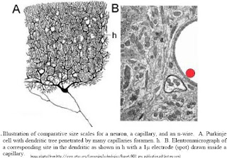
" The basic idea consists of a set of nano-wires tethered to electronics in the main catheter such that they will spread out in a “bouquet” arrangement into a particular portion of the brain’s vascular system. Such arrangement could support a very large number of probes (in the millions). Each n-wire would be used to record, very securely, electrical activity of a single or small group of neurons without invading the brain parenchyma. Obviously, the advantage of such system is that it would not interfere with either the blood flow exchange of gases or produce any type of disruption of brain activity, due to the tiny space occupied in the vascular bed.

A catheter is introduced into the femoral carotid or the sub-clavial artery and is pushed up to one of the vascular territories to be addressed. Such procedure is, in principle, similar to interventional neuro-radiology techniques where catheters are guided to any portion of the central nervous system. The number of 0.5 micron diameter wires (recording points) that could be introduced in a one-millimeter catheter is staggeringly large (in the range of few million). Once the areas to be recorded or stimulated are reached, a set of leads held inside the catheter head would be allowed to be extended and randomly distributed into the brain’s circulatory system. Since a catheter can be placed in any major brain vessels, the maximum length of nano-wire electrodes required to reach any capillary bed is of the order 2 to 3 cm. Hence, a large number of electrodes would cover any region of the central nervous system from the parent vessels harboring the stem catheters. "

This means that most of the brain can be 'reached' using this technique and signals from the 'signal ensemble in our brains' can be dynamically read to yield information on malfunctions, malformations or aberrations. This is extremely exciting and shows yet another way in which new technologies are helping us reach and probe into the deepest parts of the brain without interfering with too much of the inherent functions.

I see a future whereby patients can go a suitably equipped clinic, get this nano wire inserted into her/his femoral carotid and let the vascular system do its work of pulling the wire through all of the arteries that interlace our brains. Once the blood 'pulls' the fibers into the deepest parts of the brain, non-integrated dynamic signals from various parts of the brain can then be mixed and integrated to yield useful information helping us pinpoint tumors, clots, infarcts or damaged brain cells at a microscopic level. A neuro-surgeon can then use the same nano wire apparatus only this time turning on the ablative laser at the end of the nano-wire, find the cells that constitute the tumor/glioma and proceed to vaporize it. At the end of the procedure the nanowires are extracted out of the patients femoral carotid and the patient walks back home. No skull splitting brain surgery or no post operative recuperation... Of course this is me dreaming in the New Year...


That said, I would also urge you to refer to the following words of caution:
Release of nano-particles should be restricted due to the potential effects on environment and human health.” – Nanotechnology and Regulation within the framework of the Precautionary Principle. Final Report for ITRE Committee of the European Parliament, February 2004.

Until more is known about their environmental impact we are keen that the release of nanoparticles and nanotubes in the environment is avoided as far as possible. Specifically we recommend as a precautionary measure that factories and research laboratories treat manufactured nanoparticles and nanotubes as if they were hazardous waster streams and that the use of free nanoparticles in environmental applications such as remediation of groundwater by prohibited.” – Royal Society and Royal Academy of Engineering, “Nanoscience and Nanotechnologies: Opportunities and uncertainties,” July 2004.

Some papers of interest:

Inferring the Dynamics of “Hidden” Neurons from Electrophysiological Recordings by Valeri A. Makarov and Nazareth P. Castellanos

Fabrication of nanoelectrodes for neurophysiology: cathodic electrophoretic paint insulation and focused ion beam milling by Yi Qiao, JieChen, Xiaoli Guo, Donald Cantrell, Rodney Ruoff
and John Troy

Wiring the Brain at the Nanoscale

It Binds, Therefore I Am! by Rüdiger Vaas a review of Rodolfo Llinás’ ‘I of the Vortex’

Neuro-vascular central nervous recording/stimulating system: Using nanotechnology probes
by Rodolfo R. Llinás1 , Kerry D. Walton, Masayuki Nakao, Ian Hunter and Patrick A. Anquetil
Risk governance for nanotechnology by M.C. Roco - some very good slides on what to expect in the next generation