I remember being struck by a picture of Isabella Rossellini in which she holds her impossibly long neck proudly to the camera and also remember deciding immediately to capture this pose in oil. It took about 20 days to finish this one mainly because I was agonizing over the right colors to use for the hair. I finally decided on a combination between alizarin crimson hue, cadmium yellow pale, umber and shades of flesh for the hair. The face was mostly mixing white with a pale yellow hue contrasted with lemon yellow shade. I was happy for the most part with the results achieved mainly because it did capture the pose in the original picture (I think...). I decided to call it ‘Long neck and adornments’. The size is 48 inches by 36 inches.
Tuesday, January 30, 2007
I remember being struck by a picture of Isabella Rossellini in which she holds her impossibly long neck proudly to the camera and also remember deciding immediately to capture this pose in oil. It took about 20 days to finish this one mainly because I was agonizing over the right colors to use for the hair. I finally decided on a combination between alizarin crimson hue, cadmium yellow pale, umber and shades of flesh for the hair. The face was mostly mixing white with a pale yellow hue contrasted with lemon yellow shade. I was happy for the most part with the results achieved mainly because it did capture the pose in the original picture (I think...). I decided to call it ‘Long neck and adornments’. The size is 48 inches by 36 inches.
Friday, January 26, 2007
Damage to a brain structure called the Insula disrupts addiction to cigarette smoking:Scientists have reported recently that damage to a portion of the brain called the insula kills the urge to smoke. The insula is a region that lies underneath the cerebral cortex behind the upper lobes of our ears. It is a deep brain structure and is thought to the implicated in the intermeshing of physical activity and emotion and the translation of one to the other (including the attendant effects of motivation and craving for the ‘fix’). This may be the first time that scientists have obtained definitive proof that the insula may be the seat and originator for cravings that develop within all of us. Of course we cannot go ahead and damage a person’s insula in the hope that the person may quit smoking, but we could develop alternative therapies like selective ablation of certain portions of the insula or stem cell based mediated growth (or death) to relevant portions of the insula to stop the urge to smoke. All of that is conjecture at this point in time and only a lot more concentrated research in this field will prove to us the viability of technologies outlined above...
Dr. Antoine Bechara of the University of Southern California who led the research said that he was initially puzzled when a patient with stroke related damage to the insula suddenly gave up the smoking habit. This prompted him to look at stroke registries for correlation between location of the stroke related injuries in the brain and the predilection of the person to go back to smoking. This led to the ultimate pinpointing of the region on the brain that kills the urge in an individual to smoke. The rest they say is concentrated research and history... He has published his results in today’s issue of the science magazine... Unfortunately I have not been able to get my hands on the paper.
In light of the enormous amount of research that needs to be carried out in this area, it may be a while until we see troops of smokers heading to the operating table for brain surgery to quit smoking... Meanwhile if you want to quit smoking, I would say, just drop the bad habit.
Wednesday, January 24, 2007
Listening to the 'State of the Union', the rebuttal and the 'pre-rebuttal', I was struck by the seemingly lackadaisical attitude towards securing healthcare - especially for the indigent population.
Imagine this scenario: You belong to the middle to lower income rung of the society where you make a living from paycheck to paycheck. You are relaxing inside your home enjoying a good family get-together when you decide to get up and help yourself to some more water from the refrigerator. On your way you happen to trip on the furniture and fell down hard on your knees and double up in excruciating pain. You decide the pain is too much for you to bear and it almost seems like a broken kneecap. Your significant other manages to reach the phone, call 911 and the ambulance comes along. As you are loaded onto the ambulance they ask you about the insurance and you tell them that you are uninsured. They give you disapproving looks, but haul you off to the nearest hospital. Once in the hospital, they say that they cannot start treatment on you until you present them with a major credit card such that they could bill you for expenses. You tell them that in addition to having no health insurance (because your employer did not provide for it); you really do not have any major credit cards. They tell you to sign a few forms that will give you a credit card account immediately. You reluctantly sign and head off into the ER to be treated for a broken kneecap.
Although this scenario is made up - times are coming upon us where we would begin to see this as a form of reality among the more indigent among us. A report released this week titled "Borrowing to stay healthy" compiled by Demos (a public policy group in New York) stated that about 29% of low and middle income families with credit card debt reported using their credit cards to pay for medical expenses for major medical problems. This additional indebtedness tacked onto late fees and interest payments only adds to the suffering endured by the patient and their families.
I was also deeply struck by the following lines from the New York Times op-ed page that talked about this report:
"Its one thing to reach for your Visa or MasterCard to pay for a Barbie doll or a flat-screen TV. It’s way different to pull out the plastic because you have just learned you have cancer or heart disease, and you don’t have any other way to pay for treatment that would prevent a premature trip to the great beyond. A society is seriously out of whack when legalized loan sharks are encouraged to close in on those who are broke and desperately ill"
The report cites the following policy recommendations:
-Differentiate Medical Debt from Consumer Debt
-Limit the Entry of Medical Providers into Financial Services
-Increase Oversight of Lines of Credit Attached to Health Savings Account Products
-Improve screening for eligibility in public or private financial assistance programs
-Enact a Borrower’s Security Act
The report is a little dense reading and may not serve your light reading needs, but is nonetheless an urgent wake up call...
Read it here if time permits...
Emma Lazarus' ditty to the Statue of Liberty is worth remembering here (actually these are the last four lines of her poem 'The New Colossus' :
"Give me your tired, your poor,
Your huddled masses yearning to breathe free,
The wretched refuse of your teeming shore.
Send these, the homeless, tempest-tost to me,
I lift my lamp beside the golden door!"
Friday, January 19, 2007
The dynamics of concussion

Dr. Bennet Omalu (the neuropathologist on the case) of the University of Pittsburgh, has determined that Mr. Waters’s brain tissue degenerated into a brain that had characteristics of an 85-year-old man with early-stage Alzheimer’s disease. A lot of credit should be given to Mr. Chris Nowinski, a former Harvard football player and professional wrestler whose repeated concussions ended his career. These injuries also left him with severe migraines and depression. It also pushed him into exposing the dangers behind concussions to the brain tissue suffered from contact sports that often go unattended because in most cases the player just runs back into the field in response to cheers of heroism.
All of this got me thinking a bit more about the exact neurology behind concussion.. I would be interested in the exact neurobiology behind brain concussion, could not find too much on the web, but here is a little theory behind the sequence of events that might occur:
Our brain is suspended within our skull in a fluid medium called the cerebrospinal fluid (in fact our brain actually floats in this fluid and would our brains would limply lie on its sides without this support from the cerebrospinal fluid and the surrounding skull to give it shape and support). A concussion is trauma suffered to brain tissue usually as a result of a very sharp blow to some part of the skull that could cause temporary cognitive deficits. Sharp blows essentially translate to forces on our brain that propel the brain-mass to impact the inside of our skulls with very high velocities. High velocity brain matter can’t really go anywhere; it is abruptly stopped in its tracks by the inner surface of our skulls. In being stopped, very high impact forces are transferred from the hard dura that covers the inner surface of our skulls to the cerebral surface of the brain tissue. Sudden buildup of pressure in a localized area of the brain translates to even higher pressures inside the miniature capillaries that snake and find their way through the cerebral tissue. As brain tissue (neurons and glia) squeezes on the capillary, the blood really has no place to go but rupture the capillary and flow out in multiple minute locations (all of this might be happening at the level of minutest capillary – but nonetheless is a possibility). Of course, the sum effects of this are so small that normally nothing really happens. But repeated injuries taking place over a period of time (say over a players career), could lead to multiple blood vessel ruptures in various areas of the brain (doctors tend to call these vascular infarcts). Over time, decreased blood supplies (albeit in small quantities) and microscopic ruptures compromise the individual cells that make up our brain – the neurons. In fact necrotizing tissue could also be formed in small pockets all over the brain from the minute ruptures. Large scale cognitive deficits start to show on the individual’s personality as multiple areas involved in sensory processing and higher order executive functions start to function at decreased processing power. Some of this could be manifested as depression, migraines, headaches, listlessness etc. Maybe this is what killed Mr. Waters - we will never know for sure, but the time has come for us to look closely at the business of sending our kids off to play football or other contact sports.
I am not saying that contact sports is a bad thing, but better research needs to be dedicated to protecting the soft matter that we implicate for the development of our 'selves' within our heads.
Some of the research that I managed to dig up in this regard is here:
Traumatic brain injury and concussion in sports - James P. Kelly MD JAMA Sept 8 1999 Volume 282 No 10 Page 989
The nature of concussion: a speculative hypothesis - Paul McCrory (Br. J. Sports Med. 2001;35;146-147)
Attentional deficits in concussion - P. VAN DONKELAAR, J. LANGAN, E. RODRIGUEZ, A. DREW, C. HALTERMAN, L. R. OSTERNIG, & L.-S. CHOU Department of Human Physiology and Institute of Neuroscience, University of Oregon, Eugene, OR, USA
Essential Information for Athletes, Parents, and Coaches
Wednesday, January 17, 2007
It is always difficult to see the aftermath of drugs on human beings. The following site lists the faces of people after they have been on methamphetamine for months or a couple of years. The effect of this quiet and silent killer is stark on these faces and is being played out in a lot of homes and communities all over the United States.
- Some papers of interest:
The brains response to Methamphetamine - article
Methamphetamine dependence and HIV infection - article
Implications of research for treatment : Methamphtamine - article
Methamphetamine Dependence Is Associated With NeurocognitiveImpairment in the Initial Phases of Abstinence by Ari D. Kalechstein, Ph.D. Thomas F. Newton, M.D. Michael Green, Ph.D.
Neuropsychological Effects of Chronic Methamphetamine Useon Neurotransmitters and Cognition: A Review by Thomas E. Nordahl, M.D., Ph.D. Ruth Salo, Ph.D. Martin Leamon, M.D.
Saturday, January 13, 2007
Painting post - deviation from usual:Friday, January 12, 2007
A personal recollection and some neurological ruminations on glossolalia
Researchers at UPenn have published a neuro-imaging study on glossolalia, and have found the following:
1. Decreased activity in the frontal lobes (entirely in line with the experience – the frontal lobes maintain our sense of conscious control and thinking and the phenomenon of glossolalia shows a marked reduction of intentional control leading to low levels of activity in these lobes )
2. Decreased activity in the left hemispheric structures (our left lateral hemisphere specifically the left temporal lobe behind your ears is dominant in about 90% of individuals as they go about the mechanics of meaningful language construction, but in a ‘glossolaliac’s’ case the language is really not produced, the sounds are non-structured and non-semantic and hence lesser activity in the left lateral hemisphere)
3. The studies also showed a dip in the activity of a region called the left caudate. The caudate area is involved in motor and emotional control, so it may be that practitioners, while mindful of their circumstances, cede some control over their bodies and emotions.
4. They also noticed a shift in thalamic activity when the subject changed from a normal singing mode to a glossolaliac phase. The thalamus is responsible for relaying large amounts of cortical and cortical-subcortical neuronal information. This shift might be indicative of the sense of control alteration in which practitioners no longer feel as if they are willfully making the vocalization.
It is also interesting to note the following from this paper (more of a psychiatric treatment to this phenomena) :
Please see this paper for some more interesting related stuff:
Meditation States and Traits: EEG, ERP, and Neuroimaging Studies by B. Rael Cahn and John Polich
Thursday, January 11, 2007
Helping the blind see (device nearing end of clinical trials):I have always wondered, what if a device were invented that could input visual stimuli and multiplex it into appropriate signals and feed this as input into the optic nerve of a blind person? After the signals travel down their visual pathways into the visual areas of the brain would the blind person start to 'see'?
Well, it turns out that a company in Europe is nearing the end of clinical trials into exactly such a device (albeit a simpler version) and would be applying for licenses to commercially sell such a device in the market in early 2008.
The company is Intelligent Medical Implants AG, based in Zug, Switzerland--with its subsidiaries IIP-Technologies GmbH and Intelligent Medical Implants Ltd. (collectively referred to as "IMI Group") and is developing the Learning Retinal Implant System(TM), the first product of its developing neuro-prosthetics technology platform.
About the Learning Retinal Implant System(TM)
IMI's Learning Retinal Implant System(TM) replaces the signal-processing functions of a healthy retina and provides input to the retinal nerve cells (the ganglion cells) that, in turn, provide input to the optic nerve and the brain. The System comprises three main components: (1) an implant, "The Retinal Stimulator", which is surgically placed into the eye of a patient, who wears (2) a pair of spectacles containing an integrated mini-camera and transmitter components for wireless signal and energy transmission ("The Visual Interface"). Via a cable, the spectacles are connected to (3) "The Pocket Processor" worn at the patient's waist. This device replaces the information processing function of the formally healthy retina. The use of a high-speed digital signal processor allows the provision of "intelligent information" to the implant (and the nerve cells) by using tuneable software to approximate the information processing normally carried out by the healthy retina. The entire process enables patients to optimize their visual perception during the learning phase. Indeed, using the patient's feedback on perception as an input for the tuning of The Pocket Processor is the unique, patent-protected feature of the System and constitutes the 'learning' capability of the Learning Retinal Implant System(TM).
"The provoked visual perceptions were pleasant, according to the patients, and this was the first time they had seen something in many years-- in one case, several decades. Understandably, they reacted emotionally to their visual experience."
Looks like the Science magazine had a report on this some time back.
Monday, January 08, 2007
I did my masters in nanotechnology and every once in a while I go on the net to see what is current out there I am pleasantly surprised. I was happy to run into an article that sits at the intersection of nanotechnology and neuroscience.
Well, maybe I should backtrack and explain what nanotechnology is all about... Fortunately, I do not have to recite in detail all of the copious amount of information that is available on the net about nanotechnology, but the following article in The New Atlantis gives a very good account of what nanotechnology is all about.. (In a sentence, the human hair is about 80000 nanometers thick - we are talking about devices of the range of 10-20 nanometers thick whereas a red blood cell is vast in comparison: about 5,000 nm in diameter). At this ‘nanoscopic’ level, most of our body cells start to look like giant hot air balloons and a lot of work can be accomplished if we load the right scientific instruments at the end of nanometer sized problems that can then be inserted into our cells.
OK, this is precisely what is happening:
As you know, our brain is richly impregnated by millions of blood vessels whose diameter range from 5 millimeters (the larger junctions) to 10 microns (at the smallest levels). In fact we have about 25 kilometers of blood vessels that have a diameter of less than 10 microns.
" The basic idea consists of a set of nano-wires tethered to electronics in the main catheter such that they will spread out in a “bouquet” arrangement into a particular portion of the brain’s vascular system. Such arrangement could support a very large number of probes (in the millions). Each n-wire would be used to record, very securely, electrical activity of a single or small group of neurons without invading the brain parenchyma. Obviously, the advantage of such system is that it would not interfere with either the blood flow exchange of gases or produce any type of disruption of brain activity, due to the tiny space occupied in the vascular bed.
A catheter is introduced into the femoral carotid or the sub-clavial artery and is pushed up to one of the vascular territories to be addressed. Such procedure is, in principle, similar to interventional neuro-radiology techniques where catheters are guided to any portion of the central nervous system. The number of 0.5 micron diameter wires (recording points) that could be introduced in a one-millimeter catheter is staggeringly large (in the range of few million). Once the areas to be recorded or stimulated are reached, a set of leads held inside the catheter head would be allowed to be extended and randomly distributed into the brain’s circulatory system. Since a catheter can be placed in any major brain vessels, the maximum length of nano-wire electrodes required to reach any capillary bed is of the order 2 to 3 cm. Hence, a large number of electrodes would cover any region of the central nervous system from the parent vessels harboring the stem catheters. "
This means that most of the brain can be 'reached' using this technique and signals from the 'signal ensemble in our brains' can be dynamically read to yield information on malfunctions, malformations or aberrations. This is extremely exciting and shows yet another way in which new technologies are helping us reach and probe into the deepest parts of the brain without interfering with too much of the inherent functions.
I see a future whereby patients can go a suitably equipped clinic, get this nano wire inserted into her/his femoral carotid and let the vascular system do its work of pulling the wire through all of the arteries that interlace our brains. Once the blood 'pulls' the fibers into the deepest parts of the brain, non-integrated dynamic signals from various parts of the brain can then be mixed and integrated to yield useful information helping us pinpoint tumors, clots, infarcts or damaged brain cells at a microscopic level. A neuro-surgeon can then use the same nano wire apparatus only this time turning on the ablative laser at the end of the nano-wire, find the cells that constitute the tumor/glioma and proceed to vaporize it. At the end of the procedure the nanowires are extracted out of the patients femoral carotid and the patient walks back home. No skull splitting brain surgery or no post operative recuperation... Of course this is me dreaming in the New Year...
That said, I would also urge you to refer to the following words of caution:
“Release of nano-particles should be restricted due to the potential effects on environment and human health.” – Nanotechnology and Regulation within the framework of the Precautionary Principle. Final Report for ITRE Committee of the European Parliament, February 2004.
“Until more is known about their environmental impact we are keen that the release of nanoparticles and nanotubes in the environment is avoided as far as possible. Specifically we recommend as a precautionary measure that factories and research laboratories treat manufactured nanoparticles and nanotubes as if they were hazardous waster streams and that the use of free nanoparticles in environmental applications such as remediation of groundwater by prohibited.” – Royal Society and Royal Academy of Engineering, “Nanoscience and Nanotechnologies: Opportunities and uncertainties,” July 2004.
Some papers of interest:
Fabrication of nanoelectrodes for neurophysiology: cathodic electrophoretic paint insulation and focused ion beam milling by Yi Qiao, JieChen, Xiaoli Guo, Donald Cantrell, Rodney Ruoff
and John Troy
Wiring the Brain at the Nanoscale
It Binds, Therefore I Am! by Rüdiger Vaas a review of Rodolfo Llinás’ ‘I of the Vortex’
Neuro-vascular central nervous recording/stimulating system: Using nanotechnology probes
by Rodolfo R. Llinás1 , Kerry D. Walton, Masayuki Nakao, Ian Hunter and Patrick A. Anquetil
Sunday, December 31, 2006
Painting post - last one for this year - and a happy new year on that note:This did not come out as well as I wished it would, but this oil titled 'Hazy Shades of Winter' is 48 inches by 36 inches. I finished it in about four days and maybe that explains why parts of it are a bit sloppy (I was not the happiest with this one)... You tell me....
Friday, December 29, 2006
When opposites become apposite...In my view, science is the ultimate unifier - looking relentlessly for universal truths that explain phenomena with simple clarifying concepts. Art on the other hand is in the other extreme - the epitome of self expression, uniqueness and individuality. It is indeed refreshing when the both come together. OK, what am I talking about now…
These two worlds came together in a refreshing fusion this week at Princeton University.
Art of Science asked the Princeton University community to submit images including videos and audio produced in the course of research or incorporating tools and concepts from science. Here are the results.
Quite amazing, I must say!!
Thursday, December 28, 2006
Sobering column that appears everyday in the New York Times (for the last four years) this was from today's Times:
Tuesday, December 26, 2006
The teen brain - a work in progress and driving implications :An overwhelming amount of research is slowly bringing to light the fact that the teen brain is very much a work in progress and a lot of the 'neuronal sculpting and pruning' that takes place as the child matures through the teen years is incomplete at least until the age of 21 to 22... I remember as a teen (about 20 years back), I used to sneak into a lower shelf in my family cupboard and 'help myself' to money kept there to to buy cool jeans. For some reason my father did not confront me, but one day the goose that laid the golden eggs stopped replenishing itself and I often wondered if my father 'noticed'. I also remember sneaking out on my father’s motorcycle a couple of times without his knowledge and without any sort of a valid license to drive. I was 15 then. Looking back in retrospect, it seems stupid and unreasonable to assume that my father would not 'notice'. Research can now conclusively tell us that the teen brain is undergoing a huge amount of structural changes. In fact the following have been conclusively proved:
1. The first areas to mature (e.g., extreme front and back of the brain) are those with the most basic functions, such as processing the senses and movement.
2. Areas involved in spatial orientation and language (parietal lobes) follow.
3. Areas with more advanced functions -- integrating information from the senses, reasoning and other "executive" functions (prefrontal cortex) - mature last.
The actions outlined in 3 above takes place post puberty and extends over 8-10 years (until they are 21 or 22)... In fact actions that are directed by the pre-frontal cortex involve critical integrative functions like organize plans, generate ideas, set priorities, form strategies, control impulses, and allocate attention. In retrospect all of this was what I lacked when I helped myself to get the jeans and drove outside on my motorcycle in unbridled frivolity.
In fact a couple of months back, there was the case of a teen who texted her suicide notes as she drove her Mercedes into a oncoming Daewoo. The mother of three in the Daewoo died as a result while the teen survived. Highly irrational behavior like this can be rationalized a bit more when we understand that Louise Egan Brunstad's (later charged with murder as an adult) neuronal connections were not sculpted or pruned enough for her to think or plan or strategize her next course of actions in light of her current conditions. Of course, this opens up a bunch of ethical arguments again - do not blame the child, blame the brain - but nonetheless brings to light the fact that we need to pay closer attention to teen brains in formative modes and maybe legislate that actions that require a surprisingly large amount of planning and thinking - like driving for example should maybe be the purview of 21 year olds instead of 16-18 year old...
Of course, some of this might be a little extreme, but Louise’s case above and a bunch of other similar cases may have a case in point.
Related papers of interest:
The Adolescent Brain: A Work in Progress - A good article on changes going on in the teen brain with a focus on preventing teen pregnancy
What’s Going On In There? How the Brain and Mind Develop in the First Five Years of Life By Lise Eliot, Ph.D.
What Makes Teens Tick? - Article in TIME on the teen brain...
Cruel and Unusual Punishment: The Juvenile Death Penalty Adolescence, Brain Development and Legal Culpability - A good flyer put out by the American Bar Association that talks about law and the teen brain and the ramifications
Neuronal Regulation: A Mechanism For Synaptic Pruning During Brain Maturation by Gal Chechik and Isaac Meilijson
- a little mathematical, but a good read nevertheless
Are Teens Driving Safer? By Pilar S. Marin and Brett V. Brown, Ph.D, Child Trends
The First Three Years of Life and the Early Adolescent: Influences of Biology and Behavior - Implications for Child Rearing by Donald E. Greydanus, MD; Helen D. Pratt, PhD; Dilip R. Patel, MD - A CME review article
Teen driving - should states impose tougher restrictions?
Saturday, December 23, 2006
What you see is not reality in this really impressive optical illusion...
http://www.patmedia.net/marklevinson/cool/cool_illusion.html
"If your eyes follow the movement of the rotating pink dot, you will only see one color, pink. If you stare at the black + in the center, the moving dot turns to green. Now, concentrate on the black + in the center of the picture. After a short period of time, all the pink dots will slowly disappear, and you will only see a green dot rotating if you're lucky! It's amazing how our brain works. There really is no green dot, and the pink ones really don't disappear. This should be proof enough, we don't always see what we think we see."
The following site gives a lot more optical illusions that fool our brains if you are really into this kind of stuff... http://www.michaelbach.de/ot/index.html
Tuesday, December 19, 2006
Monday, December 18, 2006
The painting is titled 'Buy Me Bring Me Take Me'.
Thursday, December 14, 2006
Congenital AVMs and the United States Senate: Normally, arteries carry blood containing oxygen from the heart to the brain, and veins carry blood with less oxygen away from the brain and back to the heart. When an arteriovenous malformation (AVM) occurs, a tangle of blood vessels in the brain or on its surface bypasses normal brain tissue and directly diverts blood from the arteries to the veins. It is estimated that about one in 200–500 people may have an AVM and more common in males than women. Brain AVMs are usually congenital but not hereditary (meaning you may be born with one but will not pass it onto your children)...
As a brain AVM contains abnormal (“weakened”) blood vessels, they sometimes dilate over time and may eventually burst from the high pressure of blood flow from the arteries causing bleeding into the brain. The bursting may be precipitated by several factors that include exercise, stress or sometimes something as common as a sudden stop while riding in a car…
Most AVMs are detected on either a computed tomography (CT) brain scan or with a magnetic resonance imaging (MRI) brain scan. These AVMs can be surgically removed if bleeding occurs. The other ways of treating this would be to 'scar' part of the blood vessel supplying the AVM (using a technique called stereotactic radiosurgery) and allowing the AVM to 'clot off'. There are other advanced ways of treating this inclusing the usage of catheters inside the blood vessels and deploying certain specialized materials to block off the blood flowing into the AVM. Not too sure which one of this was performed on the senator..
It is interesting to note that the senator showed signs of speech slurring and had to be taken to the hospital following that. This would indicate that the AVM was somewhere in the left anterior parietotemporal lobe (just my layman theory) ;-)
Wednesday, December 13, 2006

If there is one person on a photo blog that I follow closely, it is stoneth on flickr. He compiles images and stories about poverty in the US, bittersweet most of the time...
Tuesday, December 12, 2006
Possible early detection for Alzheimer’s??Researchers at New York’s Weill Cornell Medical College found discovered certain biomarkers (a substance whose detection in our bodies indicates a particular disease state (for example, the presence of an antibody may indicate an infection)) that might seem to herald a new early test for Alzheimer’s (much before the actual onset of this devastating disease).
Researchers at Cornell seem to have found out a set of 23 proteins in the cerebrospinal fluid that seem to be an indicator for the onset of Alzheimer’s.
"Scientists believe that Alzheimer’s begins its insidious brain attack years, even decades, before forgetfulness appears — and if so, there should be evidence of those changes in the spinal fluid"
"Using a technology called proteomics, they simultaneously examined 2,000 proteins found in the spinal fluid of 34 people who died with autopsy-proven Alzheimer’s, comparing it to the spinal fluid of 34 non-demented people.
What emerged were 23 proteins, many that by themselves had never been linked to Alzheimer’s but that together formed a fingerprint of the disease."
All the patient will be required to undergo will be a spinal tap and they could be diagnosed as to whether they are at the risk of contracting Alzheimer’s later in life.
Of course a lot of this research will need to be conclusively proven, but I think that this is a quite a start for us and has a lot of potential...
Note:
"There is no single, comprehensive diagnostic test for Alzheimer's disease. Instead, doctors rule out other conditions through a process of elimination. They usually conduct physical, psychological and neurological exams and take a thorough medical history. Diagnosis is about 90 percent accurate, but the only way to confirm it is through autopsy."
"There is no medical treatment currently available to cure or stop the progression of Alzheimer's disease. There are currently five FDA-approved Alzheimer's drugs – Namenda, Cognex, Aricept, Exelon and Reminyl -- that may temporarily relieve some symptoms of the disease. Several other drugs are in development."
Wednesday, December 06, 2006
As I read more topics and books on neuroscience, the more I realize that we have only scratched the surface in understanding what really goes on in our heads. This led me to look at various sources across the web in compiling a list of five principle strategic areas that we need to focus to understand at a deeper level the activities of the ‘wet web’ that is within our heads. With this in mind, I am proposing the following five broad areas of research that universities and governments across the world will need to invest time, brains and money for achieving a better understanding in neuroscience.
1. Experience and environment driven modification of genes in the neural realm
Research and investments into genetic modifications due to neural activity in response to life events, environment, culture, beliefs, preferences and intentions across demographic groups such as family, community, society and nations. The impact of religion, ethnicity and race would be an additional orthogonal factor to the effects of the above and will need to be studied as well. This initiative will need to be funded and coordinated across multiple universities across the world.
2. Functional understanding of neural substrates spatially
Investments and research into understanding the functional aspects of different parts or 'spaces' in the human brain. Being able to zoom in or out to understand, display and explain activity in response to intrinsic and extrinsic stimulation at different levels - the synapse, the neuron, the cortical column, ensembles of cortical columns, circuits of cortical ensembles and so on until we are able to functionally explain activities across the whole brain in response to stimulus. This kind of a coordinated multi-level integration and formulation needs to be directed across various universities and hospitals. Investments in imaging technologies are crucial and the only way this will go forward.
3. Functional understanding of neural substrates temporally
Research and investments into collecting neural modifications over time. Coordinated research into clarifying changes that happen at microscopic levels like individual synapses to macroscopic levels like modifications in coordinated neural circuits that can be 'snapshotted' temporally over the lifetime of study subjects. This will yield valuable information among others on ageing, development, neurogenesis, neural substrate atrophy and temporal modification of neural activity due to disease onset/progression. This is time consuming expensive research that could span the average lifetime of a human and should be started as soon as possible
4. Databasing the brain across multiple realms - neuroinformatics
None of the above comes to fruition without extensive investments and programs in neuroinformatics. In fact there is little hope of making progress in the spatial and temporal domains described above unless we build up data warehouses that can be tapped across high speed networks by researchers across the world. This will require new university initiatives or retooling of existing informatics programs and network protocols to equip and position ourselves into providing for tomorrow’s neuroinformatics data explosion. Tools that help correlate, slice and integrate information across various data stores and common protocols and those that marry disparate data formats will also have to be studied and implemented.
5. Neurobiological basis to understanding the mind and neuroethics
Formulating a unified theory to defining the mind through the integration of various neurobiological research areas. Extensive research needs to focus on the various ‘neurobiologies’ – among others, I may quote here are the neurobiology of the senses, feeling, abstract thought, rationality, emotions and thinking culminating in a unified neurobiological understanding of consciousness. This can be achieved only by integrating results from all of the above strategies into a coherent whole that will allow us to better define our mind and self. Congruent with this research will be investments made into understanding and defining neuroethics and clearly defining boundaries for the mind and brain and legislating the definitions of brain death and mental illness in a more substantive way.
References:
1. Antonio Rosa Damasio MD, PhD
- Descartes' Error: Emotion, Reason and the Human Brain
2. Antonio M. Battro MD
- Half a brain is enough
3. Huda Akil PhD
- Mind Brain and Neuroscience
4. Vilayanur S. Ramachandran MD, PhD
- The perception of phantom limbs: The D. O. Hebb lecture. Brain, 121, 1603-1630.
5. James L. McClelland
- Retrieving general and specific information from stored knowledge of specifics: Proceedings of the Third Annual Meeting of the Cognitive Science Society, 170-172.
6. Alexander Romanovich Luria
- The Mind of a Mnemonist: A Little Book About A Vast Memory
7. Patricia S. Churchland and Terrence J. Sejnowski
- A Critique of Pure Vision
Tuesday, December 05, 2006
Painting Post
As always, I have painted a face - following my belief that the human face frames and communicates the essence of all emotions, concealed or otherwise. The size of the painting is 36 inches wide and 48 inches tall.
Monday, December 04, 2006
Brain computer interfaces: New hope for paralyzed individuals with mostly intact brain functions:25-year-old Matthew Nagle changed the channels on his TV, adjusted the volume and read an e-mail. These feats may not sound impressive, but they earned him a spot on the cover of the July 13, 2006 issue of the journal Nature (and caused a media uproar) because Nagle is a quadriplegic, paralyzed in 2001 by a knife wound that severed his spinal cord. Thanks to a system called BrainGate, Nagle was able to manipulate the TV controls, as well as a prosthetic hand, using his thoughts alone.
This company called Braingate (developed by John Donoghue, head of the Brain Science Program at Brown University, through a company he co-founded: Cyberkinetics Neurotechnology Systems) is piloting a device called Neural Interface System which consists of attaching tiny electrodes to parts of the motor strip in the brain which decodes the ensuing electrical activity into purported actions that the device wearer 'wants' to do. A dumbed down way of saying this would be that the device actually reads your thoughts.
What it is really doing is that the computer connected to the device decodes the 'motor intentions' of the individual. The electrical activity can then be used by the computer to move or align objects in the immediate periphery of the individual. Of course these objects will need to have some kind of a connection to the computer and the computer will need to go through several learning sessions to correlate certain patterns of neural electrical activity into planned goals on behalf of the individual.
"The system is designed to restore functionality for a limited, immobile group of severely motor-impaired individuals. It is expected that people using the BrainGate™ System will employ a personal computer as the gateway to a range of self-directed activities. These activities may extend beyond typical computer functions (e.g., communication) to include the control of objects in the environment such as a telephone, a television and lights."
For people living with paralysis, the technology has the potential to be life-changing.
A little more info on the implant:
The implant is a square silicon chip just four millimeters (about 1/6 of an inch) wide, studded with an array of 100 hair-thin electrodes. The chip sits on the surface of the motor cortex, while the electrodes delve midway into the two-millimeter-thick cortex to eavesdrop on neurons that normally signal muscles to move. A bundle of gold wires sends those signals out through a connector affixed to the top of the skull, and to an amplifier; they then travel by fiber-optic cable to a set of computers. During training sessions for BrainGate, the computer software learns to associate patterns of neural activity with the intent to move a hand in a particular direction; it can use those intentions to pilot a computer cursor or, if all goes as planned, a motorized wheelchair.
Think of a future where severely paralyzed people will be surrounded by intelligent robots that will feed them, clean them and cook for them exactly the way they wanted it - all through reading the thoughts of the individual.
Friday, December 01, 2006
Thursday, November 30, 2006

"The phrase "chemo brain" has come to be used by cancer survivors to describe
changes in memory, attention, concentration, and abilities to perform various
mental tasks that are associated with receiving chemotherapy treatments for
cancer. In neurology the effects are referred to as cognitive deficits or
declining neuropsychological functioning."
They have also found out the structural changes discovered are essentially reversible after some period of time. This means that after certain forms of chemotherapy, some patients could go into a "chemobrain type syndrome mode" for some time and then 'cognitively return' back to normal in time.
"They found that at one-year, patients treated with chemotherapy had smaller
volumes in cognitively sensitive areas, such as the prefrontal, parahippocampal
and cingulate gyri, and precuneus regions. However, at three-years post-surgery
there was no volume differences. That there were no differences between cancer
patients and healthy controls at any time point demonstrates that there is no
observable cancer-effect in cognitive deficits."
There is a growing hypothesis that the powerful drugs used to fight cancer are capable of diffusing through the blood-brain barrier, a protective sheath that keeps most harmful substances out of the brain. Many chemo drugs are made up of molecules small enough to breach the barrier it is thought. The publishers of this study have not made it clear that the breach of the blood-brain barrier could be the reason behind the structural changes seen in brain MRI before and after chemotherapy.
The study is published in the 1 January 2007 issue of CANCER, a peer-reviewed journal of the American Cancer Society. The following link give you a little more detail.

"We found a decrease in blood circulation in some areas of the brain in
young adults who just started to use ecstasy," said Maartje de Win, M.D.,
radiology resident at the Academic Medical Center at the University of Amsterdam in the Netherlands. "In addition, we found a relative decrease in verbal memory
performance in ecstasy users compared to non-users."
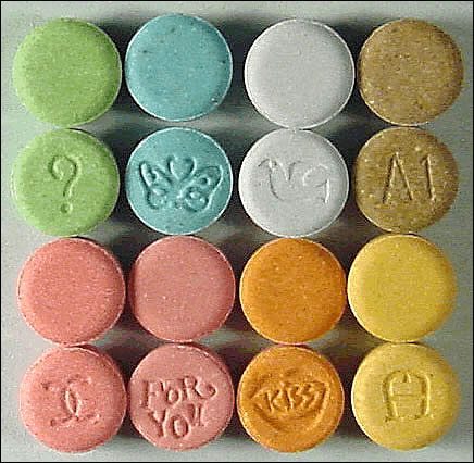
It is fairly well known that long-term or heavy ecstasy use can damage serotonergic neurons and cause depression, anxiety, confusion, difficulty sleeping and decrease in memory. However, no previous studies have looked at the effects of low doses of the drug on first-time users.
This means that even one time use of this drug is strictly a no-no if you want to look after your brain...
Wednesday, November 29, 2006

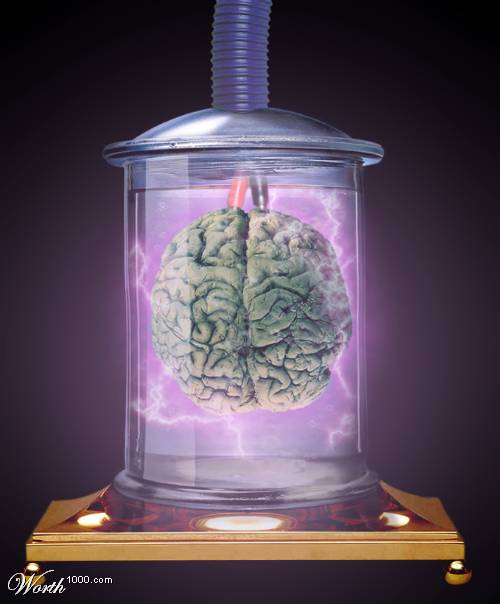
I was browsing through an online Photoshop art contest and I came upon the category called "Humans In One Million Years". I looked at this one picture and immediately thought of the following lines by Rama (V.S. RAMACHANDRAN, a neuroscientist, is professor and director, Center for Brain and Cognition UCSD).
You can browse all of the entries under the category here or browse the entire collection here…
“Lets advance to a point of time where we know everything there is to know about the intricate circuitry and functioning of the human brain. With this knowledge, it would be possible for a neuroscientist to isolate your brain in a vat of nutrients and keep it alive and healthy indefinitely.
Utilizing thousands of electrodes and appropriate patterns of electrical stimulation, the scientist makes your brain think and feel that it's experiencing actual life events. The simulation is perfect and includes a sense of time and planning for the future. The brain doesn't know that its experiences, its entire life, are not real.
Further assume that the scientist can make your brain "think" and experience being a combination of Einstein, Mark Spitz, Bill Gates, Hugh Heffner, and Gandhi, while at the same time preserving your own deeply personal memories and identity (there's nothing in contemporary brain science that forbids such a scenario). The mad neuroscientist then gives you a choice. You can either be this incredible, deliriously happy being floating forever in the vat or be your real self, more or less like you are now (for the sake of argument we will further assume that you are basically a happy and contended person, not a starving pheasant). Which of the two would you pick?”
The complete text of Rama’s article is here with some comments…
Monday, November 27, 2006

The following paper is an excellent review of visual consciousness (Highly recommended).
Just as we were carving up our turkeys and putting on the finishing touches to our Thanksgiving meals on Thursday last week, bombs carved up and put finishing touches on about 150 human beings and injured another 200 over in Iraq (all this happened in a single city - of course scores of people died in other areas also...). The stated reason for the war was that we were going in to find weapons of mass destruction.
Wednesday, November 22, 2006
I refer to this site sometime when I get bored with the usual and they have some pretty good neurology cases with complete differential diagnosis, discussion and references.
This months case seems a bit dicey and am not even too sure if this is a dyed in wool neurology case or a case of edema in the lower extremities caused by too much of sedentary activities (the patient is a computer programmer)...
Read on more about this case here. They will post the diagnosis over the month end.
Tuesday, November 21, 2006

Painting Post:The following is an oil I did entitled 'Mugshots on grilled cheese sandwiches - new trends in religiosity'. I completed this in about a month early this year.
Again, like most of my paintings, this one is 48 inches long and 36 inches wide. This is meant to be a commentary on the current trend of finding religious icons in grilled cheese sandwiches and selling the same on e-bay for thousands of dollars... What were the seller and buyer thinking? Reminds me of a fad that happened about 10 years ago in India when stone idols of Ganesha (a Hindu god) suddenly started drinking milk from devotees cups... This sure is an opium for the masses...
Beneath thin veneers of cultivated sensibility lies raging undercurrents of pent up bigotry held in check by social pressures and lawsuits. OK, what am I spouting here now???
I am a big fan of the 'Kramer' character in the 'Seinfeld' show, but a recent tirade by Mr. Michael Richards onstage that both forth in full resplendence his innate feelings towards ingrained dispositional representations of skin color is worth mentioning here. The video of the incident is even more shocking and it really shows what a lot of ordinary (looking) folks around us have on their minds and how it all comes spewing out when they are pushed just little bit. In this case all that was required to trigger this amazing dispensation was a heckler in the crowd.
Monday, November 20, 2006
Out of Body experiences triggered by electrical simulation of the angular gyrus
Yet another notch for science. We have all read about mystical sounding abnormal 'out of body' experiences where people seem to inhabit a non-corporeal self and seem to look down upon their own bodies from a position outside their body. Rational explanation for a lot of this could not be unearthed and it was thought that either these people were faking it or these people were closer to some superior force. Some people were just waiting for a rational scientific explanation. Looks like the people who were waiting just got it...
Dr. Olaf Blanke, a neurologist at the École Polytechnique Fédérale de Lausanne in Switzerland reported in the Sept. 21 issue of Nature magazine of cases of women who sensed a shadow person behind her on electrical simulation to certain parts of the brain.
As we know the angular gyrus in the temporal parietal junction forms one of the higher association areas for senses like vision, sound and touch. Located behind the somatosensory strip and ventral to the primary visual areas with inputs from the speech and sound areas, it is possible that this area could provide a representation for our 'self' in a rudimentary form (in the same sense as one gets a feeling for a 'phantom limb'). On electrical simulation to this area, people have reported seeing or feeling detached from the corporeal self and have also found to experience intriguing behavior.
"There is nothing mystical about these ghostly experiences, said Peter Brugger, a neuroscientist at University Hospital in Zurich, who was not involved in the experiments but is an expert on phantom limbs, the sensation of still feeling a limb that has been amputated, and other mind-bending phenomena.
“The research shows that the self can be detached from the body and can live a phantom existence on its own, as in an out-of-body experience, or it can be felt outside of personal space, as in a sense of a presence,” Dr. Brugger said. "
The woman replied that she had a weird sensation that another person was lying beneath her on the bed. The figure, she said, felt like a “shadow” that did not speak or move; it was young, more like a man than a woman, and it wanted to interfere with her.
When the current was turned off, the woman stopped looking to the right, and said the strange presence had gone away. Each time he reapplied the current, she once again turned her head to try to see the shadow figure.
When the woman sat up, leaned forward and hugged her knees, she said that she felt as if the shadow man was also sitting and that he was clasping her in his arms. She said it felt unpleasant. When she held a card in her right hand, she reported that the shadow figure tried to take it from her. “He doesn’t want me to read,” she said.
Because the presence closely mimicked the patient’s body posture and position, the doctor performing the test concluded that the patient was experiencing an unusual perception of her own body, as a double. But for reasons that scientists have not been able to explain, he said, she did not recognize that it was her own body she was sensing.
Read on more here.

Finally I managed to photograph some of my oils over the weekend and am planning on slowly posting them up here.. I am not the greatest at photography. I laid out the painting on a wooden deck on our backyard and took a snapshot (tried my best to not take the wooden rafters, but in a couple of cases they do show through)...
The following was an oil I did entitled 'Yearning - A tribute to Leonardo'. I completed this in about a month in November of last year.
The painting is about 48 inches long and 36 inches wide.
Friday, November 17, 2006
As I attempt to continue my armchair explorations into neurology, I am slowly realizing that unless I have a good idea of the various neural substrates and their spatial existence within our heads, it may be very difficult for me to understand where the structures are in the first place and secondly, how they are interconnected. With this in mind, I modified an elegantly laid out thought experiment in one of the books that I was reading (Neuroanatomy through Clinical Cases by Hal Blumental from Yale) and thought that you might enjoy this piece.
(It might be useful to use this diagram as a guide as you go through your journey)
It is basically a thought experiment where I am a scuba diver with a powerful flashlight on a mission to find the hippocampus (that elusive organ that is supposed to orchestrate our short and long term memories. The same thought experiment allows me to miniaturize myself such that I would be able to pass myself though a syringe used for a lumbar puncture on one of my close friend Jack who has volunteered to sit through my explorations. Included in my survival kit is also a miniature copy of Grey’s anatomy that will help me navigate my way through some of those tough sounding Latin named structures that I am going to encounter along the way – hey, we all need a Rand McNally’s when we go on a long exciting trip, don’t we? So here goes…
OK, that minitaturization hurt a little, but I was happy to pass easily through the lumbar puncture syringe into the subarachnoid space of Jack’s lumbar cistern passing through the skin, subcutaneous tissues, the hard interspinous ligament, through the tough dura mater and finally into the cerebrospinal fluid in the subarachnoid space. As soon as I was released into the cerebrospinal fluid, I stretched my legs and hands and looked around me to try and learn what was going on… Instinctively I notice that I was placed above the vertebral body S1 between L3 and L4. I also notice that I am bounded internally by the pia mater and externally by the arachnoid. As I start to swim in the cerebrospinal fluid, I start to notice a lot of wispy spiderweb like protrusions called the arachnoid trabecula from the outer arachnoid wall that seem to swim and sway in the cerebrospinal fluid and some of them even reach out and touch the pia mater. Carefully avoiding the spiderweb like protrusions, I use my flippers to swim upwards a little bit. I look up and I see wondrous rope like filaments descending all around I am somehow reminded of a horse’s tail. The tail seems to sway in the cerebrospinal fluid all the way into the lumbar cistern. I understand that I am looking at the cauda equina. Looking upwards and in front of me, I see a gleaming whitish pink tube like organ through the translucent pia and look at the conus medullaris portion of the spinal cord. I swim around the cord a little bit trying my best to avoid all of the horse tail like strings and see some of the nerve roots entering and exiting the spinal cord. The ones entering the spinal cord seem to be sensory nerves from the dorsal side (Jack’s back side) and motor nerves seem to be exiting on the ventral side of the whitish cord.
I make my way up, swimming against the flow of the cerebrospinal fluid for quite a while until I see a large opening up above me. It seems to be about the level of Jack’s mouth and realize that I am looking at the foramen magnum (a large ring shaped entrance to the cranial cavity). It is a bit foreboding as I seem to be heading into a large chamber of some sort and realize that this is the Cisterna Magna. I look up and see the ventral aspects of the cerebellum that looks like grayish pink structure with lots of little goose bumps all over… Below me I see that the spinal cord has given way to become the medulla. I also realize that the reticular formation forms some part of the floor beneath me if I stand here… I swim around the whitish pink cord and am fascinated by looking at the pons on the opposite side. I also seem to have fallen into the Pontine cistern on the other side. I quickly extricate myself from the Pontine cistern and come back to the cisterna magna and notice that there is another ventricular structure around me to either side. On either side of me, I can see the lateral foramina of Luschka. In fact I can slide down the walls of this foramen, but decide against it as I have more interesting things up there and let’s not forget, I am in search of the hippocampus. I decide to swim up against the dorsal side of the medulla and as soon as I come up a little above the cerebellum I notice the midline foramen of Megendie. The cerebellum still stretches on above me… a pulsating greyish mass that seems to be busy calculating coordination, gait and other higher motor functions. Without stopping at the foramen of Megendie, I decide to keep swimming upstairs… The force of the cerebrospinal fluid seems to be especially strong now and I force myself upward and notice that I have entered a large cavity and realize that this is the fourth ventricle that I am in…. I stop swimming and slowly land on the ventral floor of the fourth ventricle. Rostrally, I see the pons up ahead and caudally, I see the medulla. You must realize that I am standing perpendicular to Jack’s upright posture. (if I wanted to shoot out of his body, I would come head first out of the lower portion of his head at this time – just to give you a visual of where I was) Dorsally, the roof seems to be the cerebellum, with the large cerebellar peduncles on either side. I look at the miniature copy of the Grey’s anatomy that I am carrying and notice that the floor is also called the rhomboid fossa (containing important structures like the facial colliculus and the sulcus limitans). Parts of the floor rostrally also seem to be colored bluish grey and on looking it up is the locus ceruleus, which owes its color to an underlying patch of deeply pigmented nerve cells, termed the substantia ferruginea. I turn my headlight again rostrally and put it on high beam and realize that now I have come to a very narrow passage that I doubt that I will be able to cross. I start swimming upwards and with great difficulty clamber my way through a narrow tunnel called the cerebral aqueduct of Sylvius. The rush of the cerebrospinal fluid at this portion is really strong and I had to use all of my energy in staying my course at this point in time. I swam some of the way and walked some of the way as the tunnel seems to gradually slope with an upward trajectory. I also notice that there is a substantial grey matter at this point in time and slowly understand that I am crossing the periaqueductal grey portion of the midbrain (an important descending pathway which can be instrumental in inhibiting pain signals from the spinal cord). Clambering out of the tunnel, I thought that I could relax for a bit, but I seem to be immediately pulled down and start to sink to the depths of another cavity – the third ventricle. As I am traveling down the third ventricle, I look to my left and right and notice that I first pass the walls of the thalamus and then the walls of the hypothalamus. The third ventricle seem be bounded by the thalamus and hypothalamus on the left and the right. I also notice that the two thalami seem to be joined tighter at the interthalamic adhesion midway along the third ventricle.
At this point I stop swimming, look up and notice two parallel white arches running along the roof of the ventricle and realize that I am looking at parts of the fornix running over me. This is really exciting! I also notice that there is a profusion of capillary like vessels separated from the subarachnoid space by pia mater. Liquid seems to be filtering through ependymal cells (a type of neuroglia) from blood to become the cerebrospinal fluid that I am swimming in. I now realize that I was seeing this all along my way except along the narrow walls of the cerebral aqueduct, but only now did I begin to notice this. Looks like the cerebrospinal fluid is being made all along the ventricular system. I look behind me and notice the pineal and the suprapineal recesses on the caudal end of the third ventricle. I decide not to go the caudal end, but I seem to have two ways to go forward or rostrally, I can either swim upwards entering one of the two narrow tunnels that I see there or I can swim forward (rostrally) the third ventricle and see if there is anything out there that will lead me to the hippocampus. I first decide to swim straight ahead (rostrally). No luck. I seem to have come up against a dead end of some sort, but I notice that I can touch the supra optic recess (above the optic chiasma – where Jack’s optic nerves birfucate), and the infundibular recess (above the pituitary stalk). I know that the hypophysis is somewhere at the end of the infundibular recess, but I have other things to do… I also notice that walls of the hypothalamus extend all its way down here also. Well, now that I have had no luck, I decide to swim back up and squeeze myself through the tunnel to my right. On consulting Grey’s I find that I am entering the right foramen of Monro. Just as I am entering this new tunnel, I pause and look around me and I notice that I am standing on the anterior commissure (a bundle of white fibers, connecting the two cerebral hemispheres across the middle line) with my left hand up on the fornix and my right hand on the walls of the thalamus. I swim my way up this passage and find myself in a larger chamber – in fact one of the largest chamber that I have been in so far. Well, this is it – it is the right lateral ventricle. I start to swim forward in this cavity to find my way around a little better and I reach the rostral or anterior end of the lateral ventricle. This is called the anterior horn. Looking up, I see a bunch of white fibers running in close formation and recognize that as the corpus callosum – that great highway of fibers that connect the two hemispheres. I also realize that I am deep in the frontal lobes of Jack’s head. The floor here seems to be the head of the caudate nucleus.
I then decide to turn all the way around and swim to the other ends of this great chamber and realize that in reality this big cavity is made of three large horns, the anterior or the frontal horn that I just ran into, the posterior or the occipital horn and the inferior or temporal horn located lower down in the temporal lobes. I am sure that I will be able to find the hippocampal structures somewhere here. As I swim back I feel like I am being sucked into the foramen on Monro and have to swim quite strong against the current of the cerebrospinal fluid that is draining into the third ventricle. I seem to be in the body of the lateral ventricle now… I look to my right and see a translucent wall of membrane called the septum pellucidum. The septum pellucidum is located in the midline of the brain, between the two cerebral hemispheres. It is attached superiorly (above), anteriorly (in front), and inferiorly (below) to the corpus callosum, the large collection of nerve fibers that connect the two hemispheres. Inferiorly and posteriorly (in back), it is attached to the anterior part of the fornix. I shine my headlight through the septum pellucidum and look into the further reaches of Jack’s left lateral ventricle. To my left, I see a huge grey mass bulging into the walls of the lateral ventricle and realize that this is the body of the caudate nucleus. I also notice that I am able to make out the outlines of part of the thalamus, the choroids plexus and the fornix from my vantage point in the body of the lateral ventricle. I decide to keep swimming forward and presently find myself in the posterior horn of the lateral ventricle. Now I am in close proximity of the occipital lobe but still no sign of the hippocampus. I still notice that on looking up, I can see the great fibers of the corpus callosum running silently overhead carrying all of that important information between the hemispheres. I reach a dead end here also and then turn right around and am planning on heading back home when I lose my step and fall headlong into a long curvy descending passageway down and slowly realize that I am falling down the steep slope of the temporal horn of the lateral ventricle.
Luckily I do not hurt myself and manage to gather myself and stand up and look around. Upwards I see nothing but the white of the sky and realize that I am staring at Jack’s right cerebral hemisphere inside the temporal lobe – the seat of his higher thinking and memory. Anteriorly, along the median, I see the stria terminalis. The stria terminalis extends from the region of the interventricular foramen to the temporal horn of the lateral ventricle, carrying fibers from the amygdala to the septal, hypothalamic, and thalamic areas of the brain. It also carries fibers projecting from these areas back to the amygdala. It participates in anxiety and stress responses. I also notice that I can see the tail of the caudate nucleus upfront. I turn my head caudally and notice the amydaliod nucleus towards the terminal end of the inferior horn. OK, now where is the hippocampus. I look down and cannot seem to believe my eye.. There in a row I recognize the fimbria (a prominent band of white fibers along the medial edge of the hippocampus) the hippocampus itself and the collateral eminence (elevation along the floor of the posterior part of the temporal horn of the lateral ventricle, lateral to the hippocampus caused by the deep collateral sulcus). I realize at this point that I am standing on the hippocampus!!!!
Now I need to find the shortest way out – but that is another story.